Thorax Phantom
Katia Sourbelle
The aim of this phantom is to provide semi–anthropomorphic cone–beam rawdata for CT-examinations of the thorax.
Phantom Description
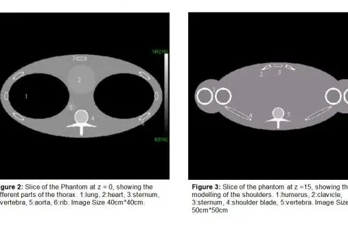
The Phantom should represent the important parts of the thorax which are usually visualised during the CT examination : lungs, heart, aorta, ribs, spine, sternum, and shoulders.
Thorax:
The lungs are represented by two ellipsoids and the heart by a sphere. The aorta consists of three joined cylinders (2 vertical and 1 horizontal), to represent its ascending and descending parts.
The ribs will be modelled by nine small joined cylinders (Fig 1.b). In order to define the cylinders, we will say that their center lies on a tilted ellipse with the co-ordinates {a cos(q ), b sin(q ), c sin(q )}, for q between p /2 and 3p /2 with steps of p /10. The axes of the cylinders are parallel to the tangent of the ellipse at the centers.
We will take 5*2 ribs for the whole thorax.
For the spine we use the model defined for the Cardio-Phantom (Stefan Ulzheimer, IMP) which will be built to evaluate the quality of CT images (Fig 1.c).
The Sternum will be represented by a box.

Shoulders:
The clavicle is represented by a cylinder and the shoulder blade by the half of an ellipse. In order to simulate the usual stark attenuation at the shoulders, we will modell them by two spheres.
All bones are filled with marrow. The whole Phantom will be made of 271 basic objects. The exact definition of the phantom are given in the following tables. The units are cm for the lengths and g/cm3 for the densities. The relevant objects lie between z=-15 cm and z=+18 cm. The field of view is 50 cm.
Files
