Translational Molecular Imaging
- Imaging and Radiooncology

Prof. Dr. Leif Schröder
Abteilungsleitung Translationale Molekulare Bildgebung
The research of the Division of Translational Molecular Imaging focuses on the development and characterization of targeted contrast agents in the context of novel detection techniques for diagnostic applications in oncology. We aim to visualize important molecular changes in the occurrence and progression of cancer at an early stage and benefit from the strong interdisciplinary tradition of magnetic resonance in Heidelberg.
Image: Illustration credits: Barth van Rossum, FMP.,
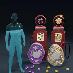
Image: Illustration credits: Barth van Rossum, FMP.,
Our Research
To this end, we are pursuing the approach of translating current scientific concepts to diagnostics. These include novel magnetic resonance methods such as spin hyperpolarization and saturation transfer in exchange-coupled spin systems. We also investigate bioscientific aspects of established contrast agents with regard to their long-term stability and expand the understanding of their biochemical behavior.
Following its establishment with the support by the Dieter Morszeck Foundation, the divisionbegan its work in spring 2023 in the newly installed Joint NMR Facility (jointly operated with the Department of Drug Discovery) and has a strong connection to the Faculty of Physics and Astronomy at Heidelberg University due to its scientific focus. The techniques we have developed are also used in various exploratory studies with international cooperation partners. The paradigm shift pursued in our laboratory aims to combine the advantages of non-invasive imaging such as MRI with the high sensitivity and specificity of functionalized reporters. This is a crucial building block of early detection and therapy monitoring and will eventually close the gap between nuclear medicine modalities such as PET / SPECT and MRI without using ionizing radiation or compromising on penetration depth as with optical methods. A key aspect of this is to fundamentally re-address the low sensitivity of conventional MRI at the molecular level and thus make molecular markers and processes that were previously impossible to visualize visible in MRI.
Spin physics for ultra-sensitive MR imaging
New approaches for better diagnostics
Conventional magnetic resonance imaging (MRI) is relatively insensitive and therefore usually only shows the signals of fat and water molecules. However, it has the advantage of being able to do without ionizing radiation and to show deeper tissue with excellent soft tissue contrast. Until now, however, tumors have often only been detected when they exceed a certain size and already contain billions of cells. The combination with contrast agents, which visualize biochemical changes such as the metabolism of the tumour or cellular markers at a much earlier stage, has therefore been a long-standing goal in MRI research.
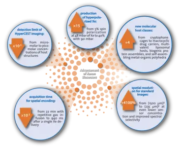
A central component of this is to increase the sensitivity of MRI. Translation from physics is used here to exploit the quantum mechanical properties of nuclear spins in order to achieve increased magnetization for a 10,000-fold increase in signal. In combination with molecular biotechnology methods, we integrate these spin systems into reporters that target cancer-specific markers. Our methods are based in particular on the harmless noble gas xenon, which can be put into a state of extremely high magnetization using powerful lasers. The methods we have developed have already increased the hyperpolarization of xenon for these applications by a factor of 15 and reduced the detection limit by a factor of 100,000. Overall, we can visualize target molecules in images after 100 s, where conventional MRI would otherwise take 1100 years.
Since the spin systems in living tissue only retain the previously prepared state for a limited time, it is also of central importance to understand the physical principles of these processes and to further optimize the reporters. A paradigm shift in the generation, delivery and encoding of spin magnetization is crucial in order to use it for new diagnostic methods.
Selected projects
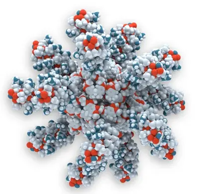
Koselleck project multivalent host structures
The addressing of target molecules by molecular units, which are then combined as reporters with hyperpolarized atoms in situ only after their binding to cells in a second step, enormously expands the range of applications of hyperpolarized MRI. In this interdisciplinary project, which is funded by the DFG's highly endowed Koselleck Programme, novel nanocarriers are being developed that can accommodate a large number of hyperpolarized atoms in the tissue. More info
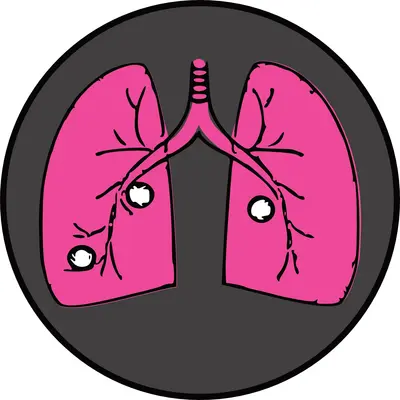
WWCR project on molecular breast cancer MRI reporter
With the support of the World Wide Cancer Research (WWCR) foundation, we are developing a new class of molecular lung MRI reporters that are combined with the inert gas xenon. Xenon as a gas is well suited for detecting breast cancer metastases in the lungs by simple inhalation. With this project, we are striving for a new type of personalized diagnostics in MRI for the early detection of metastases. More info

Stability of Gd contrast agents
Gadolinium-based contrast agents (GBCAs) can interact with endogenous substances under certain conditions. Our division contributes new methods to the understanding of these interactions in several collaborations. More info
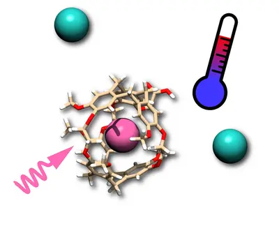
BIOQIC project for MR-based temperature measurement
MRI measurements using the (hyper)CEST method apply a large number of radio frequency pulses. This can lead to temperature fluctuations in the sample and change the image contrast. This project of the DFG-funded graduate school BIOQIC (GRK 2260) investigates such effects with hyperpolarized Xe, which can act as both CEST reporter and temperature probe. More info
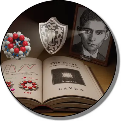
Advanced k-space encoding for hyperpolarized spins
The encoding of spatial image information for hyperpolarized spins poses particular challenges due to the limited lifetime of hyperpolarization. Various approaches can be utilized to avoid redundant information and to improve image quality with reduced acquisition times during both acquisition and image reconstruction. More info
Team
- Show profile

Prof. Dr. Leif Schröder
Abteilungsleitung Translationale Molekulare Bildgebung
- Show profile

Sandra Casula
Administrative Assistant for Divisions E041, E270 and E280
-
Viktoria Bayer
-
Hannah Gerbeth
-
Dr. Jabadurai Jayapaul
-
Luca Kempny
-

Dr. Alexandra Lipka
Group leader
-
Samuel Lehr
-
Dr. Patrick Werner
Postdoc Biologie
Selected publications
(highlighted as cover article) Jost JO, Schröder L
Jayapaul J, Komulainen S, Zhivonitko VV, Mareš J, Giri C, Rissanen K, Lantto P, Telkki V-V, Schröder L:
Werner P, Taupitz M, Schröder L, Schünke P
(highlighted as cover article) Kunth M, Schröder L
Schröder L, Lowery TJ, Hilty C, Wemmer DE, Pines A
Methods
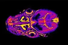
MRI experiments are performed on a 400 MHz extra-wide-bore NMR magnet with an Avance Neo console. The gradient system is capable of achieving amplitudes of 150 G/cm. Different coils for proton and X-nucleus detection are available. More info
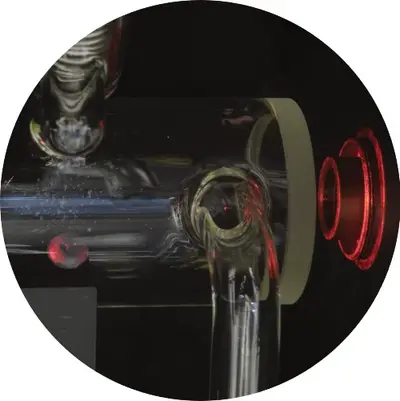
The production of hyperpolarized xenon is based on Spin Exchange Optical Pumping (SEOP) and is carried out with our specially developed LEIPNIX polarizer (Laser Enabled Increase of Polarization for Nuclei of Imprisoned Xenon). More Info
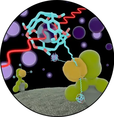
To develop sensitive measurement techniques for xenon biosensors, we use an indirect detection scheme of the transiently bound xenon. This which utilizes the exchange of the noble gas between the free state in solution and the biosensor molecule: chemical exchange saturation transfer with hyperpolarized nuclei. This achieves a signal amplification of about 7-8 orders of magnitude. More info
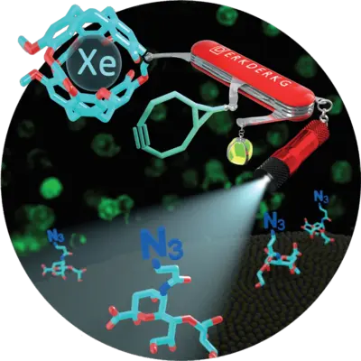
NMR signals of the noble gas xenon react extremely sensitively to their molecular environment. In order to utilize this molecular specificity for NMR contrast agents, xenon can be temporarily enclosed in molecular cages that bind specifically to a certain target molecule. More info

The quantification of the influence of paramagnetic ions on the magnetization of water protons allows conclusions to be drawn about the interaction of macromolecules such as glycosaminoglycans (GAGs) with these ions, which are used for MR contrast agents. More info
Teaching and theses

Winter semester:
(planned from 2025: NMR Physics of Coupled Spin Systems)
Summer semester:
Medical Physics 2 (together with PD Dr. Kuder)
Joint events together with the Divison of Medical Physics in Radiology

Winter semester:
Methods of Physics in Biology and Medicine (together with PD Dr. Kuder and Prof. Dr. Seco)
Co-organizer of the DKFZ Medical Physics Seminar
Summer semester:
Methods of Physics in Biology and Medicine (together with PD Dr. Kuder)
Joint events together with the Divison of Medical Physics in Radiology
Our department regularly offers opportunities for project internships on various NMR-relevant topics. We are constantly updating a list of project options, which may later be used to develop final theses. Under certain conditions, the internships can also be credited as part of the advanced internship of the Faculty of Physics and Astronomy.
Students can gain insights into the interdisciplinary field of MR research and learn various experimental techniques of nuclear magnetic resonance as well as aspects of image reconstruction and processing. A brief overview of topics can be found here.
Contact via leif.schroeder(at)dkfz.de
Topics for Bachelor's and Master's theses as well as doctoral projects can be assigned to interested parties after consultation. A brief overview of topics can be found here. Previous knowledge of medical physics or MR physics from the corresponding curriculum of the Faculty of Physics and Astronomy is advantageous. Applications for doctoral projects are usually submitted centrally via the DKFZ PhD program (link).

Our department, in collaboration with Prof. Meldrum, organizes a recurring, multi-week visit by student groups as part of the "Study Abroad" program of the College of William & Mary in Virginia.
Exemplary cooperations and joint projects
In the field of hyperpolarized MR and especially within the HYPERBOLIC consortium there is a close collaboration with the NMR spectroscopy and CEST imaging group.
The Joint NMR Facility in Technology Park 4 (INF 581) is operated together with Dr. Aubry Miller's Department of Drug Discovery. Users from various departments have access to a 400 MHz narrow-bore and a 400 MHz wide-bore magnet.
For the synthesis of new reporters, we work together with Dr. Martin Schäfer on peptide synthesis.
The validation of our reporters with regard to cell culture studies and mass spectrometry is carried out jointly with the working group of Martina Benesova-Schäfer at the Research Center for Imaging and Radiation Oncology (REZ, INF 223).
In this multi-year joint project of the German Center for Translational Cancer Research (DKTK), we are developing new methods of MRI with hyperpolarized reporters at the Heidelberg site together with partners in Freiburg, Tübingen and Munich. Our department is contributing to the expansion of applications beyond metabolic imaging in order to visualize a marker on certain cancer cells beyond the lifetime of hyperpolarized metabolites such as pyruvate.
Within the Health + Life Science Alliance Heidelberg Mannheim, we are working together with the Institute of Organic Chemistry (Prof. Mestalerz) and the Institute of Inorganic Chemistry (Prof. Enders) on new molecular cage structures that can bind xenon reversibly and exhibit improved exchange kinetics.
Some of the department's doctoral students (currently Samuel Lehr and Hannah Gerbeth) are taking part in the CancerTRAX program, in which the projects are led by a tandem of supervisors at the DKFZ and WIS. Our department cooperates with Prof. Bar-Shir's group to develop new MR reporters.
Together with the GlycoScience of CellularBiophysics working group headed by Dr. Böhm, we are developing new methods to understand the interaction of Gd(III) ions with components of the extracellular matrix.
In a collaboration with the Collaborative Research Center "Matrix in Vision", Patrick Werner uses NMR relaxometry methods to investigate the behavior of Gd(III) ions from MRI contrast agents under various biochemical stimuli.
Our department is a partner in the EU-funded "I4World" project to investigate new molecular host structures.
Further links
Get in touch with us


