Cookie Settings

We use cookies to optimize our website. These include cookies that are necessary for the operation of the site, as well as those that are only used for anonymous statistic. You can decide for yourself which categories you want to allow. Further information can be found in our data privacy protection .

Essential

These cookies are necessary to run the core functionalities of this website and cannot be disabled.

Name Webedition CMS
Purpose This cookie is required by the CMS (Content Management System) Webedition for the system to function correctly. Typically, this cookie is deleted when the browser is closed.
External media

Content from external media platforms is blocked by default. If cookies from external media are accepted, access to this content no longer requires manual consent.

Name YouTube
Purpose Show YouTube content
Name Twitter
Purpose activate Twitter Feeds

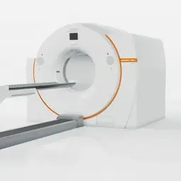
© dkfz.de

Fig1. Image of a PET scanner. The principle of positron emission tomography (PET) is the so-called coincidence measurement, i.e. simultaneous detection of two 511 keV photons emitted in 180 degrees.

PET / CT (general information)

Positron emission tomography (PET) is a nuclear medical procedure that allows noninvasive and quantitative measurement of radiopharmaceutical concentrations. Positrons produce an annihilation radiation of 511 keV, which is emitted at an angle of 180°. Based on the principle of a coincidence measurement, the reconstruction of the acquired projections takes place in transversal, sagittal and coronal slices. Furthermore, correlation to morphological imaging methods, such as CT and MRT, is simply possible by means of the new combined devices, such as PET-CT and PET-MRT.

Various biological parameters such as tissue perfusion, transport and metabolism of radioactive tracers as well as the kinetics of therapeutically active substances (e.g. cytostatics) can be measured with PET. The most commonly used tracer is the fluoro-18-labeled deoxyglucose (FDG), a glucose analogue that is enhanced in most of the tumors. F-18-deoxyglucose (FDG) is transported and phosphorylated analogously to glucose, but is then trapped and not metabolized further than 90%.


The main advantages of PET are:

  1. The use of short-lived radiopharmaceuticals, which can be applied one after the other (so-called multitracer examinations) in order to combine various biological informations about the tumor (e.g., perfusion, amino acid transport and glucose metabolism).

  2. The use of positron-emitting isotopes such as oxygen-15 (O-15), nitrogen-13 (N-13) and carbon-11 (C-11). The o.g. isotopes are more suitable for the labeling of biomolecules and pharmaceuticals since they do not alter their biological behavior.

  3. The exact, absolute measurement of radiopharmaceutical concentrations. This is a clear measurement technology advantage as compared to SPET (Single Photon Emission Tomography). The reason for this is that in the case of PET the measurement of the coincidence event is independent of the depth. Furthermore, with the aid of a transmission measurement, an exact determination of the absolute attenuation factors can be carried out for each coincidence line.

  4. Acquisition of dynamic studies as PET scanners are full-ring systems.


PET has become increasingly important in oncology, as the sensitivity of FDG-PET is good for staging and differential diagnosis. In the majority of the studies, the use of PET is limited to the purely visual interpretation of whole body images. Quantitative measurements (mostly static acquisitions) are used for most applications. A semiquantitative evaluation is partly used for therapy monitoring. This evaluation is usually carried out via the "Standardized Uptake Value", SUV. This parameter allows a comparison of patient examinations by standardizing the measured concentration for the administered dose and the body weight as the distribution volume estimate. SUV is a dimensionless distribution value, a SUV of 1.0 means equal distribution of a radiopharmaceutical.

A dynamic data acquisition is, in principle, superior to a static measurement since only the measurement of the kinetics of a tracer can provide accurate information about its temporal and spatial distribution in the target region. A differentiated analysis of the kinetic data is useful in order to obtain the optimal information from the PET examination.


Positron Emission Tomography Computed Tomography (PET-CT):

The PET scanners were replaced by the combination of a modern PET and a CT system. The CT component is used for the attenuation correction of the PET images and the assignment of the PET findings. Depending on the question, a PET measurement can be combined with a diagnostic CT examination.

Diagnostics and staging in tumors using positron emission tomography/ computed tomography (PET-CT)

a) Clinical trials to detect recurrences and metastases in patients with malignant tumors using fluorodeoxyglucose (FDG) and fluorothymidine (FLT).

The increased glucose metabolism or proliferation are used as a biomarker to improve tumor diagnosis and staging. FDG is accumulated by enhanced transport and phosphorylation in the tumor tissue that is increased in almost all tumors. The principle of tumor visualization with FLT is based on a thymidine kinase I activity which is increased during the S phase, which phosphorylates FLT and thus leads to the accumulation of the tracer.

b) Studies on receptor expression with labeled peptides, such as Ga-68-DOTATOC, Ga-68 Bombesin, Ga-68 Melanocortin (MSH), and Ga-68-PSMA.

Here, PET is used to measure receptor expression in various tumors, such as neuroendocrine tumors, gastrointestinal stromal tumors (GIST), prostate carcinomas, and melanomas. The labeled peptides can be administered in combination with fluorodeoxyglucose (FDG) in order to characterize the tumors biologically. This focus also encompasses correlative studies of tracer kinetics and molecular biology parameters (e.g. gene expression data of the tumors).

© dkfz.de

Fig. 2: Patient with a recurrent gastrointestinal stromal tumor (GIST) of the stomach and a suspicious liver lesion.
Above left: transversal PET-FDG uptake, which shows an increased metabolism in the stomach and in the right liver lobe.
Top right: CT shows the two suspicious lesions in the stomach and in the ventromedial part of the right hepatic lobe.
Bottom left: transversal Ga-68 Bombesin PET uptake showing a significantly elevated Bombesin uptake in the liver lesion and a slight Bombesin increase in the stomach. This indicates a different receptor expression. Both lesions were malignant (histologically confirmed).

© dkfz.de

Fig. 3: Patient with a Lung Tumor (Non Small Cell Lung Cancer) in the left hilus. Detection of increased FDG metabolism and increased somatostatin II receptor expression in the lung carcinoma.

© dkfz.de

Fig. 4A: Patient with a primary prostate carcinoma (Gleason score 3 + 4) in the left lobe. Detection of increased PSMA expression in prostate carcinoma. The results are recorded in the fused PET-MRT (upper left) and in the fused PET-CT (lower left).
The Ga-68-PSMA time activity curve (right) above the tumor (blue curve) shows a continuous increase of the tracer.

© dkfz.de

Fig. 4B: Patient with a metastatic prostate carcinoma. The maximum intensity projection (MIP) PET images (left) after injection of Ga-68-PSMA show multiple metastases (bone, lung, lymph node metastases). The fused PET-CT images (right) allow a precise anatomical assignment of these metastases.

Therapy follow-up studies in tumors for the assessment of the response to chemotherapy using positron emission tomography/computed tomography (PET-CT)

PET with FDG is a sensitive method that can be used for therapy monitoring. For the evaluation of PET data sets, a semi-quantitative evaluation is carried out based on the so-called "Standardized Uptake Value" (SUV). SUV is a value representing the global uptake of a radiopharmaceutical at a defined time. In order to estimate the long-term response to chemotherapy, we have investigated an integrated evaluation of tracer kinetics, which takes into account the transport rates and the distribution volume of FDG. The significance of this evaluation with regard to the detection of long-term therapeutic effects and the assessment of individual survival is examined in the following tumors:

a) Non-Small-Cell Lung Cancer (NSCLC)

FDG follow-up examinations are performed before, after one and three cycles of chemotherapy with vinorelbine and oxaliplatin.


b) Colorectal carcinomas

FDG follow-up examinations are carried out before, after and after four cycles of chemotherapy with FOLFOX (fluorouracil, folinic acid, oxaliplatin).


c) Multiple myelomas

FDG follow-up examinations are carried out before, after one and three cycles of anthracycline-containing chemotherapy (e.g. VAD).


d) Soft tissue sarcoma

FDG follow-up examinations are performed before, after one cycle and preoperatively following induction therapy with AIG (adriamycin, ifosfamide, growth factor) in high-risk patients with soft tissue sarcomas intended for high-dose chemotherapy with peripheral blood stem cell rescue (PBSCR).


e) Gastrointestinal stromal tumors (GIST)

PET follow-up studies are performed before and after therapy with the tyrosine kinase inhibitor imatinib.


f) Melanoma stage IV

PET follow-uo studies are performed before and after various immunotherapies (immuno-checkpoint inhibitors).

© dkfz.de

Fig. 5: PET follow-up study with F-18-FDG in a patient with a lung metastasis (right hilary) of a colorectal carcinoma. Reduction of FDG metabolism after one cycle of chemotherapy with a combined protocol (FOLFOX). The kinetic analysis of the FDG data primarily indicates a reduction in phosphorylation (k3). These data suggest a reduction in the growth rate of metastasis after chemotherapy.

© dkfz.de

Fig. 6: PET follow-up study with F-18-FDG in a patient with a gastrointestinal stromatumor (GIST) of the small intestine. Significant reduction in FDG metabolism after one month therapy with the tyrosine kinase inhibitor Imatinib as a sign of a very good therapeutic effect.

© dkfz.de

Fig. 7: PET follow-up study with F-18-FDG in a patient with metastatic melanoma. Multiple metastases predominantly pulmonary, which show no response to immunotherapy with Ipilimumab. After conversion of the therapy to Ipilimumab and Vemurafenib, clear response. The fused PET-CT images of the lung show a clear response of the lung metastases after the combined therapy.

Materials for tissue generation in systemically diseased bone (SFB-TR 79)

a) Diagnosis and staging in patients with multiple myeloma based on dynamic PET-CT (dPET-CT) with F-18 deoxyglucose (FDG) and Na-18 fluoride (fluoride)

Within the scope of this study the glucose metabolism (measured with FDG) as well as bone reconstruction (measured with fluoride) should be investigated in patients with confirmed multiple myeloma. The aim of the study is to investigate the value of the method for the detection of active myeloma lesions as well as the correlation of both tracers with stage and activity of the disease (Fig. 6). Furthermore, the significance of the parametric imaging is to be evaluated (Fig. 7).


b) Therapy in patients with multiple myeloma based on dynamic PET-CT (dPET-CT) with F-18 deoxyglucose (FDG) and Na-18 fluoride (fluoride)

Here, the response to a chemotherapy is to be recorded with dPET-CT and resistant lesions shouldbe detected, which can then be treated with local therapies.

© dkfz.de

Fig. 8: Example of a PET-CT examination with F-18 deoxyglucose (FDG) and Na-18 fluoride (fluoride) in two patients with multiple myeloma. On the left side is a patient with a recurrent myeloma, who shows an increase in the FDG metabolism in a rib on the left side. Several bone lesions are delineated in the fluoride PET, since bone metabolism is also increased in fractures and degenerative lesions. On the right side is a patient with a primary myeloma, who has several hypermetabolic lesions in FDG, which are indicative for myeloma manifestations. The corresponding fluoride images of the bone metabolism show only a few lesions with increased bone metabolism. FDG has so far proven to be a suitable tracer for the detection of active myeloma manifestations.

© dkfz.de

Fig. 9: Top right: FDG cross-section in small pelvis in a patient with multiple myeloma.
Bottom right: Parametric image showing the phosphorylated part of FDG. Due to the higher contrast the delineation of the lesions is better.

to top
powered by webEdition CMS