7 Tesla MRI: Proton imaging and RF pulse design
7 Tesla MRI: Proton imaging and RF pulse design

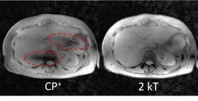
The research group focuses on MR proton imaging in humans at ultrahigh field (UHF) of 7 Tesla with a special interest in body imaging at this field strength. Due to the short radiofrequency (RF) wavelength, the spatial distribution of the magnetic component of the RF field (B1+), which is necessary for the imaging process, is highly inhomogeneous. This yields inhomogeneous signal and contrast distributions in the body (see Figure), making a diagnosis difficult or impossible. To counteract this effect, we use multi-transmit (Tx) body coils in combination with parallel transmission (pTx) techniques and dedicated RF pulses.
The spatial variation of the B1+ field can be altered by pTx and thereby homogeneous flip angles of the proton spins can be achieved, which homogenizes the contrast. A special focus of the working group is the use of the "MRExcite" body coil (RF arrays), which, unlike conventional transmit coils, is not placed locally on the body, but is installed in the scanner behind the bore liner. This coil contains 32 Tx channels (multi-channel transmit systems) and thus offers a higher degree of freedom for pTx compared to most local coils. One of the goals of the research group is to take advantage of the system and to evaluate the benefits and challenges of 7 Tesla body imaging for future clinical applications.
Several projects in the group are performed in close collaboration with the UHF group at the Physikalisch-Technische Bundesanstalt (PTB) in Berlin.
Research Topics
- Imaging of the human body at UHF using multi-transmit channel body coils
- Development of novel pTx methods for UHF body and brain imaging, particularly when using the 32-Tx-channel "MRExcite" body coil
- Development and investigation of B1+ mapping techniques for the "MRExcite" coil regarding applicability, accuracy and precision
- Investigation of quantitative velocity imaging techniques for UHF
Clinical translation of the developed methods in patient studies

Radiofrequency (RF) arrays play a crucial role in MRI. In all modern MRI systems, arrays of RF coils are used to detect the signals emanating from the spins in the human body. Larger number of elements in such receive arrays can increase the SNR and allow for higher acceleration of the acquisition.
While clinical MRI systems at 1.5T and 3T use birdcage volume coils for transmission and arrays for reception, at ultra-high field (UHF) MRI, RF arrays are not only used for reception, but also for transmission. This is to gain more degrees of freedom when shaping the transmit field, as volume coils do not produce a homogenous excitation field at UHF. Similar to beam forming in modern radar technology, the elements of a transmit array in MRI are driven with individual amplitudes and phases and even different pulse shapes.
These arrays can be characterized be a multitude of criteria. They should have a high transmit efficiency to provide a strong excitation field with the transmit power provided by the system, as well as a high SAR efficiency to allow for a strong excitation field without damaging tissue due to excessive heating. Furthermore the elements of the array need to be well decoupled and their respective transmit profiles need to be distinct from one another to provide a high degree of freedom for shaping the transmit field.
While transmit RF arrays at UHF are almost exclusively local arrays placed directly on the body, our group aims at developing RF arrays that are integrated in the MRI system behind the inner cladding of the bore, just like the transmit volume coils of lower field MRIs [1].
Our current and future work in this area includes:
- Finding transmit antennas best suited for integrated arrays
- Developing very thin arrays to fit between boreliner and gradient coil of the most modern MRI systems
- Developing meta material solutions for improved coil performance
References: [1] Orzada, S., et al. (2019). "A 32-channel parallel transmit system add-on for 7T MRI." PLoS One 14(9): e0222452

Since the beginning of Magnetic Resonance Imaging (MRI) there has been a constant drive to higher magnetic field strengths. The reason for this is the enhanced signal-to-noise-ratio (SNR) and improved contrast the increase in main magnetic field strengths provides. While 1.5T and 3T have become clinical standard, higher fields have emerged in scientific application with 7T having recently found its way into clinical practice.
While a rise in SNR and enhanced contrast can potentially increase the diagnostic value of the images acquired with higher field strengths, higher field strengths also bring challenges. The radio frequency (RF) fields used to excite the spins have to have the same frequency as the spins. This is called the Larmor frequency and it is proportional to the main magnetic field strength. Since the wavelength of RF waves is anti-proportional to frequency, the waves become much shorter at higher main magnetic field strengths. For example at 7T the wavelength in tissue at the Larmor frequency is about 11-13cm which is smaller than the diameter of even a human head. This leads to wave effects with brighter and darker areas with in the field of view, which severely affect image quality. To cope with this, multichannel approaches can be used where multi-channel transmit arrays are driven by the multiple amplifiers of a multi-channel RF system. With such a system, the effects of the reduced wavelength on the image quality can not only be reduced, but also more sophisticated transmit techniques like selective excitation can be enhanced. Furthermore, the increased degrees of freedom provided by multichannel transmit systems allow more control over local specific absorption rate (SAR), allowing for shorter repetition times and higher flip angles.
Our group has developed and implemented a 32 channel transmit system as an add-on for the 7T MRI system of the DKFZ [1].
The system consists of 32 IQ-modulators, 32 RF 2 kW peak power amplifiers with pre-distortion, a power supervision for patient safety, and integrated RF body array and peripheral devices to control correct timings.
The high channel count and high RF power of the system allow to acquire images with a large field of view tailored excitation to get closer to the full potential of 7T MRI.
Currently we are working on:
- Modulators with higher resolution and sampling rate
- More efficient amplifiers for high duty-cycle sequences
- A more modular design to allow for even higher channel counts
References: [1] Orzada, S., et al. (2019). "A 32-channel parallel transmit system add-on for 7T MRI." PLoS One 14(9): e0222452
Contact
-
Sebastian Schmitter
Group leader
