Cookie Settings

We use cookies to optimize our website. These include cookies that are necessary for the operation of the site, as well as those that are only used for anonymous statistic. You can decide for yourself which categories you want to allow. Further information can be found in our data privacy protection .

Essential

These cookies are necessary to run the core functionalities of this website and cannot be disabled.

Name Webedition CMS
Purpose This cookie is required by the CMS (Content Management System) Webedition for the system to function correctly. Typically, this cookie is deleted when the browser is closed.
External media

Content from external media platforms is blocked by default. If cookies from external media are accepted, access to this content no longer requires manual consent.

Name YouTube
Purpose Show YouTube content
Name Twitter
Purpose activate Twitter Feeds

Publications Snapshots

Fitness trade-offs incurred by ovary-to-gut steroid signaling in Drosophila, by Ahmed SMH et al., Nature 2020

Fitness trade-offs incurred by ovary-to-gut steroid signalling in Drosophila

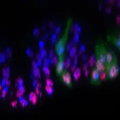
Sex hormones increase the risk of oncogenic mutations triggering tumours: Left: Intestinal stem cells with a carcinogenic mutation (green), right: the same cells under the influence of the sex hormone
© dkfz.de

Sex hormones affect homeostasis and disease progression in reproductive organs. However, it is not well understood whether this sexual dimorphism in circulating hormones can also impart differences in the physiology of non-sex organs such as the gut. Here, we show that the ovary-derived steroid hormone 20-hydroxyecdysone (20HE) in Drosophila females drives intestinal stem cell proliferation, leading to larger guts. Exposing males, which have low 20HE levels, to 20HE in their food is sufficient to instate female growth phenotypes to the gut, suggesting that the circulating levels of 20HE are a strong determinant of ISC behavior. However, this proliferation advantage normally given to female ISCs comes at the expense of faster gut deterioration during aging and higher tumor predisposition. This work provides a mechanism that connects elevated levels of sex steroids to physiology and pathology of non-sex organs. Owing to the interesting parallels between Drosophila and mammals, more detailed analysis of sex steroid effects on the human gut should be relevant and worthwhile [pubmed].

Selective 40S footprinting reveals cap-tethered ribosome scanning in human cells, Bohlen J et al., Molecular Cell 2020

Model summarizing our data of translation reinitiation

Model summarizing our data of translation reinitiation
© dkfz.de

Nearly 50% of all human mRNAs contain at least one uORF (upstream Open Reading Frame) in front of the main ORF and many of these are translated in vivo. Upon terminating uORF translation, ribosomes need to reinitiate translation on the main ORF in order to express the encoded protein. This mechanism is not well understood. When translating the uORF, most initiation factors (eIFs) are thought to fall off the ribosome. One open question is how new eIFs are re-recruited to the ribosome after uORF translation to enable translation of the main ORF downstream.

We developed a technique which we call 'selective 40S footprinting' to visualize initiating 40S ribosomes on endogenous mRNA in vivo and to reveal the positions on the mRNAs where eIFs join the ribosome and then leave again. This revealed several fundamental observations about translation initiation in human cells. Surprisingly, contrary to our expectation, we found that eIFs do not fall off the ribosome right away when it starts translating. Instead, the ribosome retains eIFs for roughly 12 elongation cycles. Hence on short uORFs, this means ribosomes are still eIF-associated when they terminate, and hence can restart a new round of translation on the same mRNA. We think selective 40S footprinting will be a useful tool to study translation initiation mechanisms in vivo on endogenous mRNAs. [pubmed]

Cdk4 and Cdk6 Couple the Cell-Cycle Machinery to Cell Growth via mTORC1, Romero-Pozuelo J et al., Cell Rep 2020

Cdk4 and Cdk6 Couple the Cell-Cycle Machinery to Cell Growth via mTORC1, Romero-Pozuelo J et al., Cell Rep 2020

© dkfz.de

Cell growth is coupled to cell-cycle progression in proliferating cells but the underlying molecular mechanisms are not well understood. It is known that CyclinD-Cdk4/6 contributes to cell cycle progression. We show now that CyclinD-Cdk4/6 simultaneously activates cell growth in mammalian cells via activation of mTORC1, the major regulator of cell growth.
Our findings also provide new insights into the mechanism of the Cdk4/6 inhibitors palbociclib and abemaciclib which are clinically used to treat breast cancer. Cdk4/6 inhibition acts on cell proliferation and growth via at least two parallel pathways: by blocking cell-cycle progression and by inhibiting mTORC1. This could provide an explanation why combined inhibition of Cdk4/6 with mTORC1 inhibitors is an effective strategy for blocking proliferation of ER-positive breast cancer cells, because cells would need to develop resistance to both drugs to reactivate proliferation. [pubmed]

Crebl2 regulates cell metabolism in muscle and liver cells, Tiebe M et al., Sci Rep 2019

Crebl2 regulates cell metabolism in muscle and liver cells

Common Crebl2 targets in myoblast and hepatoma cells. Overlap of Crebl2 dependent genes in C2C12 myoblasts and Hepa1–6 cells.
© dkfz.de

Our previous work identified a new transcription factor complex in Drosophila, Reptor and Reptor-BP (Tiebe et al., 2015). These factors are responsible for the majority of the transcriptional output of mTORC1 in Drosophila flies. Intriguingly, instead of regulating cell size as most mTORC1 effectors, Reptor and Reptor-BP strongly affected the  glucose and fat metabolism of the adult fly.
 We now extended this work into mammalian cells. Specifically, we found that loss of Crebl2, the mammalian ortholog of Reptor-BP, causes an increase in fat stores in both mouse muscle and liver cells in vitro. Crebl2 loss also causes further metabolic dysfunction in muscle cells, e.g. increases in glucose uptake, beta-oxidation rates, ATP levels and lactate secretion. Transcriptional analysis showed hundred of genes are affected by loss of Crebl2 and many genes regulated by mTORC1 are dependent on Crebl2 in mouse embryonic fibroblasts and muscle cells.
 We suspect future studies will evaluate whether Crebl2, as in flies, also regulates metabolism in mice and mediates processes downstream of mTORC1. [pubmed]

Damage sensing by a Nox-Ask1-MKK3-p38 signaling pathway mediates regeneration in the adult Drosophila midgut, Patel PH et al., Nature Communications 2019

Damage sensing by a Nox-Ask1-MKK3-p38 signaling pathway mediates regeneration in the adult Drosophila mid

This is a fly intestine under pathogenic infection. The green cells are new cells produced during intestinal regeneration. The red signal is p38 activation in intestinal epithelial cells upon damage.
© dkfz.de

This study investigates how intestinal epithelial cells sense stress or damage, defend themselves and promote epithelial regeneration. It shows that the Nox-ROS-ASK1-MKK3-p38 signaling axis integrates various stresses to promote intestinal regeneration. 
Epithelial tissues are regularly exposed to a variety of stresses caused by pathogens, toxins, and other environmental factors. Epithelial cells have an incredible ability to defend themselves from these insults and to repair themselves, but how stress sensing is achieved and then coupled to epithelial regeneration is not well understood.
The study uses the adult fly intestine, which is remarkably similar to our own intestine. The data suggest that Nox-Ask1-MKK3-p38 signaling senses multiple different stresses, including pathogenic bacterial infection and chemical and mechanical insult, in intestinal epithelial cells and effectively promotes intestinal stem cell (ISC) proliferation.
This work has potential application for regenerative medicine and diseases that develop from the loss of epithelial integrity such as inflammatory bowel diseases, which increases the risk for colorectal cancer. [pubmed]

Gene expression atlas of a developing tissue by single cell expression correlation analysis, Bageritz et al., Nature Methods 2019

Gene expression atlas of a developing tissue by single cell expression correlation analysis, Bageritz et al., Nature Methods 2019

Schematic representation of the method used to generate computed expression maps.
© dkfz.de

This study in close collaboration with the Boutros lab provides a gene expression map of the Drosophila wing disc.
The map was developed using a new method for analyzing single cell RNA sequencing data based on gene expression correlations rather than cell mapping.
The Drosophila wing disc is an important model system for studying tissue growth, pattern formation, epithelial morphogenesis, intercellular signaling, cell competition, and tissue biophysics.
The challenge with the wing disc is that it is composed of many undifferentiated, pluripotent cells. In addition, the transcriptome coverage of current single-cell RNA sequencing approaches does not allow to confidently determine the tissue location of a sequenced cell based on its expression of known genes.
We therefore developed a new method that uses a cross-correlation matrix between known mapping genes and all genes in the genome to generate expression maps. We thereby discover 824 genes with spatially restricted expression patterns. The approach identifies both known and new clusters of genes with similar expression patterns.
As proof of concept for the functional correlation of genes in a cluster, we find that the previously uncharacterized gene CG5151, which correlates with Wnt4 and wg gene expression, indeed regulates Wnt signaling.
The novel method is a useful tool for generating expression atlases of undifferentiated tissues during development. [pubmed]

Dietary stearic acid regulates mitochondria in vivo in humans, Senyilmaz-Tiebe et al., Nature Communications 2018

Dietary stearic acid regulates mitochondria in vivo in humans, Senyilmaz-Tiebe et al., Nature Communications 2018

Dietary C18:0 induces mitochondrial fusion in human neutrophils. Scheme of the clinical cross-over study. Subjects followed a low-C18:0 (vegan) diet for 2 days and then were given a banana milk shake either containing or lacking C18:0 (24 g) in a randomized fashion. Blood samples were collected at indicated time points. Each subject underwent the whole procedure twice, once with each milk shake. The study was double-blinded.
© dkfz.de

Our previous work identified the metabolite stearic acid (C18:0) as a signaling molecule that regulates mitochondrial morphology and function in fly and human cells in culture. We found that when C18:0 is added into the medium of cells in culture, this leads to activation of a signaling pathway that starts with post-translational modification of a cell surface receptor, TfR1, and leads to activation of Mitofusin 2 and mitochondrial fusion. In this paper we conducted a clinical study to test whether this pathway is also activated in humans upon eating C18:0. Indeed, we find that 3 hours after eating C18:0, the mitochondria of our white blood cells become more fused. This indicates that the human body senses and reacts to the levels of C18:0 in our diets. In contrast, palmitic acid (C16), the most abundant fatty acid in our food, does not activate a mitochondrial response like C18:0 does. Interestingly, epidemiological studies showed that C16:0 increases the risk for cancer and cardiovascular risk whereas C18:0 reduces both. This work was done in collaboration with the lab of Peter Nawroth at the Heidelberg University Hospital and was funded by an ERC Consolidator Grant. [pubmed]

This publication is in the TOP 50 Nature Communications life and biological sciences articles published in 2018.

DENR•MCTS1 heterodimerization and tRNA recruitment are required for translation reinitiation, Ahmed and Schleich et al., PLOS Biology 2018

DENR-MCTS1 heterodimerization and tRNA recruitment are required for translation reinitiation

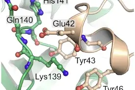
Residues involved in the interaction between DENR and MCTS1. DENR-Glu42 interacts with the nitrogen of the main chain of MCTS1-Gln140 and His141. Furthermore DENR-Tyr43 and Tyr46 hydrogen-bond with MCTS1-His141 and Lys139, respectively.
© dkfz.de

Usually eukaryotic ribosomes translate only a single Open Reading Frame on an mRNA and then dissociate from the mRNA. In some cases, when there is a short upstream Open Reading Frame (uORF) that precedes the main Open Reading Frame, ribosomes can translate the uORF, terminate translation, and then undergo a poorly understood process called ‘translation reinitiation’ whereby they resume scanning for another AUG inititation codon, and then translate the main Open Reading Frame. The molecular functions required for translation reinitiation are not known. We previously showed that two non-canonical initiation factors DENR and MCTS1 promote translation re-initiation. To get a deeper molecular understanding of how DENR and MCTS1 work, we generated a 2.1 Å high resolution crystal structure of MCTS1 bound to the DENR minimal binding domain (N terminal amino acids 24-51). This allowed us to identify individual amino acid residues in DENR and MCTS1 important for heterodimerization and for tRNA binding. Importantly, by mutating these specific amino acid residues, we discover that the DENR•MCTS1 complex needs both functions, heterodimerization and tRNA binding, to promote translation re-initiation in vivo. This thereby identifies for the first time two molecular functions needed for translation reinitiation. [pubmed]

Elevated levels of the reactive metabolite methylglyoxal recapitulate progression of Type 2 Diabetes, Moraru et al., Cell Metabolism 2018

Elevated levels of the reactive metabolite methylglyoxal recapitulate progression of Type 2 Diabetes

© dkfz.de

Several studies have shown that type 2 diabetes progresses even if patients are under optimal glycemic control. This suggests that insulin resistance and hyperglycemia are not the root causes for the disease, and raises the question of the underlying molecular changes that drive type 2 diabetes initiation and progression. Previously it was shown that type 2 diabetes patients have elevated levels of methylglyoxal (MG), a reactive metabolite that forms adducts on proteins and alters their function. In this study we find that Drosophila knockout animals that are impaired in MG detoxification have elevated endogenous MG levels and progressively developed insulin resistance, obesity and hyperglycemia, the core characteristics of type 2 diabetes. This suggests elevated MG can recapitulate some of the core aspects of diabetes in this model organism, raising the possibility that altered MG production or detoxification could be a cause of type 2 diabetes. Future work will be needed to see if this also applies to humans. [link to press release], [pubmed]

Changes in global translation elongation or initiation rates shape the proteome via the Kozak sequence, Acevedo et al., Scientific Reports 2018

kozak_sequence, Changes in global translation elongation or initiation rates shape the proteome via the Kozak sequence

The balance of translation initiation versus translation elongation determines overall translation efficiency.
© dkfz.de

The levels of individual proteins, such as transcription factors that control cell specification, or oncogenes that control cell proliferation, are critical for determining cellular phenotypes in health and disease. Protein levels are determined by a combination of mRNA synthesis, mRNA turnover, protein synthesis and protein turnover. Although work in the past focused on transcriptional regulation, recent work has highlighted that the step of translation – the conversion of mRNA to protein – is critical in determining protein levels in a cell. Molecular mechanisms exist that regulate the rate of translation of individual mRNAs, or classes of mRNAs, thereby ‘sculpting’ the cellular proteome. This study here identifies one such molecular mechanism that cells have in their ‘toolbox’ for determining the level at which proteins get made.

During eukaryotic translation initiation, the ribosome searches for an appropriate AUG start codon. This process takes into account the RNA sequence directly upstream and downstream of the AUG – the ‘Kozak sequence’. If the Kozak sequence is strong (e.g. accAUGg), there is a high likelihood that the ribosome will initiate translation at the AUG. Surprisingly, many transcripts in both humans and in Drosophila have weak Kozak sequences. This seems counter-intuitive at first because it suggests that millions of years of evolution have not selected the sequence around that AUG for efficient translation. One explanation could be that these particular proteins need to be present at low levels in cells, hence the Kozak sequence has been selected for low level expression. This would be a ‘constitutive’ effect that occurs in all cells in the organism and under all conditions. We find in this study that the Kozak sequence also plays an additional role that can cause expression of this class of weak-Kozak containing mRNAs to be differentially regulated in response to changes in cell signaling or stress. 

This paper finds that the balance of translation initiation versus translation elongation is important in determining overall translation efficiency. When mRNAs have a strong Kozak sequence, then translation initiation is efficient, and therefore translation elongation becomes rate-limiting. Hence, drops in translation elongation rates will cause mRNAs with strong Kozak sequences to become less expressed. In contrast, mRNAs with weak Kozak sequences have low initiation rates and hence translation elongation is not limiting. This means elongation rates can drop without having much effect on translation of mRNAs with weak Kozak sequences. Therefore, this identifies a mechanism whereby a ‘global’ drop in elongation rates will differentially affect translation of mRNAs with strong versus weak Kozak sequences thereby sculpting the proteome. [pubmed]

CycD/Cdk4 and discontinuities in Dpp signaling activate TORC1 in the Drosophila wing disc, Romero-Pozuelo J et al., Developmental Cell 2017

CycD/Cdk4 and discontinuities in Dpp signaling activate TORC1 in the Drosophila wing disc, Romero-Pozuelo

Cells with high TORC1 activity are in early S phase.
© dkfz.de

mTORC1 is an oncogenic kinase that powerfully drives cell growth when active. How mTORC1 activity is regulated in living tissues has not been carefully studied until now because of the lack of adequate reporters that work in situ. This paper studies for the first time the activity of mTORC1 in a growing organ during animal development using the Drosophila wing as a model system. We generate a new anti-phospho-RpS6 antibody that efficiently reports mTORC1 activity by immunostaining. Surprisingly we discovered that in the developing wing tissue, mTORC1 activity is not uniform, but patchy and controlled by the cell cycle regulator CycD/Cdk4 to peak at the G1/S transition. This indicates that CycD/Cdk4 is linking the cell cycle to the main regulator of cell growth, mTORC1. [pubmed]

DENR•MCT-1 Promotes Translation Reinitiation Downstream of uORFs to Control Tissue Growth, Schleich et al., Nature 2014

DENR•MCT-1 Promotes Translation Reinitiation Downstream of uORFs to Control Tissue Growth

DENR-KO die as pharate adults with larval-like abdominal epidermis. DENR promotes cell proliferation and boosts protein synthesis in proliferating cells.
© dkfz.de

DENR and MCTS1 are oncoproteins that interact physically with each other and have been linked to neurological disorders and to cancer. Although DENR has a protein domain suggesting a role in mRNA translation initiation, the molecular functions of DENR and MTCS1 were not known. To understand the function of DENR, we generated DENR knockout Drosophila. These knockout flies die due to impaired proliferation of abdominal epithelial cells. Interestingly the phenotypes of DENR knockout flies are not observed when general translation is impaired, suggesting DENR and MCTS1 might regulate translation of a specific subset of mRNAs involved in cell proliferation.

The analysis of mRNAs that require DENR for efficient translation revealed that DENR-MCTS1 specifically facilitate the expression of mRNAs with upstream open reading frames (uORFs) that have strong Kozak sequences (stuORFs). In Drosophila, these are roughly 200 mRNAs that code for regulatory proteins and kinases involved in cell proliferation and neurobiology.

uORFs are located in the 5´untranslated region of mRNAs (5 ´UTR). Many eukaryotic transcripts (>50% in human and mouse cells) have uORFs that precede the initiation codon of the main ORF. If the uORF has a strong Kozak sequence, the scanning ribosome (coming from the 5´ cap) will select the start codon of the uORF for translation initiation. In order to allow translation of the main downstream ORF, the ribosome needs to terminate translation of the uORF and then re-initiate translation. This process likely requires recycling of all translation initiation components and re-recruitment of an initiator tRNA. Our findings showed that DENR-MCT1 specifically promote this translation reinitiation process. These findings suggested a new control system regulating the translation of a specific set of mRNAs via DENR-MCT1. [pubmed]

Identification of transcripts with short stuORFs as targets for DENR•MCTS1-dependent translation in human cells, Schleich et al., Sci Rep 2017

DENR and MCTS1 are oncoproteins that interact physically with each other and have been linked to neurological disorders and to cancer. We previously discovered that DENR and MCTS1 promote translation re-initiation in Drosophila (see above). mRNAs containing short upstream Open Reading Frames (uORFs) with strong Kozak sequences specifically require DENR and MCTS1 for their translation. To understand how DENR and MCTS1 affect human cells, we studied here which human mRNAs require DENR and MCTS1 for their translation. We found that in human cells, DENR and MCTS2 regulate translation of mRNAs with very short stuORFs, coding for just 1 or 2 amino acids. This identified circa 100 genes as putative DENR and MCTS1 translational targets. One of the mRNAs that responds most strongly to DENR or MCTS1 knockdown is that of GPR37. GPR37 affects oligodendrocyte differentiation and myelination, as well as dopamine uptake by neurons. Mutations in GPR37 have been identified in autism patients. The identification of DENR and MCTS1 target transcripts will serve as a basis for future studies aimed at understanding the mechanistic involvement of DENR and MCTS1 in diseases like cancer and autism. [pubmed]

Oxygenation and adenosine deaminase support growth and proliferation of ex vivo cultured Drosophila wing imaginal discs, Strassburger et al., Development 2017

The Drosophila wing imaginal disc has been an important model system over the past decades for discovering novel biology related to development, signaling and epithelial morphogenesis. We discovered an explant setup that supports both growth and proliferation of wing discs for 9 hours ex vivo. We found this requires both oxygenation of the tissue and adenosine deaminase activity in the medium. This is a first step towards extending the duration of ex vivo culturing to enable in the future the live imaging of this organ as it grows and develops. [pubmed]

THADA regulates the organismal balance between energy storage and heat production, Moraru et al., Developmental Cell 2017

De novo mutations in DENR disrupt neuronal development and link congenital neurological disorders to defective mRNA translation re-initiation

Fat body of THADA KO animals stained for neutral lipids with Bodipy 493 reveals that they have more lipid per cell and larger lipid droplets compared to controls.
© dkfz.de

Thyroid Adenoma Associated (THADA) is an interesting gene for many reasons. As the name implies, it was found mutated in thyroid adenomas and several other types of cancer. Genome-wide association studies (GWAS) identified THADA as one of the top genes associated with type 2 diabetes. THADA has also played a role in human evolution. It is one of the most strongly selected genes that differentiates modern humans from Neanderthal. More recently, it was amongst the most strongly selected genes as humans adapted to warm or cold regions. Surprisingly, however, the function of THADA was not known.

Using Drosophila as a model system, we discovered that THADA regulates the organismal balance between energy storage (as fat) and energy consumption (to produce heat). THADA knockout flies are obese, but produce too little heat and are cold-sensitive. We found THADA does this by regulating the ER calcium pump SERCA. Interestingly, the obesity of THADA knock-out flies was rescued by re-introducing human THADA into the flies’ genomes, suggesting that THADA has the same metabolic effect in both humans and fruit flies. This work provides the first functional and mechanistic characterization of this fascinating gene, and it provides a link between climate adaptation and genetic predisposition for obesity in humans. [pubmed]

Evidence against a role for the Parkinsonism-associated protein DJ-1 in methylglyoxal detoxification, Pfaff et al., J Biol Chem 2016

In vivo, carbonyls react with thiol and amino groups of proteins, nucleic acids and amino lipids, leading to the formation of advanced glycation end products and protein cross-links. The resulting destruction of proper protein function puts the cells under stress. DJ1, a protein associated with Parkinsonism, was previously shown to act as a protein deglycase that repaired glycated proteins. In both DJ-1 knockdown cells in culture and in DJ-1β knock-out flies, we could detect no accumulation of protein adducts caused by methylglyoxal, and no effect on the viability of cells in response to methylglyoxal. [pubmed]

De novo mutations in DENR disrupt neuronal development and link congenital neurological disorders to defective mRNA translation re-initiation, Haas et al., Cell Reports 2016

Neurological disorders such as autism are complex phenotypes with complex underlying molecular causes. Hence the discovery of mutations and genes that are linked to such diseases is useful for improving our molecular understanding of these diseases. This work discovers a de novo mutation in the gene DENR in a patient with autism. This work shows that perturbations in DENR disrupt the migration and terminal branching of cortical neurons in the mouse brain. Based on our previous work on DENR and its partner MCTS1, we find that the DENR de novo mutation found in this patient disrupts the ability of DENR to promote translation re-initiation. [pubmed]

to top
powered by webEdition CMS