Biomedical Physics in Radiation Oncology
- Imaging and Radiooncology

Prof. Dr. Joao Seco
Head of Division of Biomedical Physics in Radiation Oncology
Radiotherapy (RT) plays a key role in the treatment of numerous solid tumors. It involves the precise targeting of high-energy X-ray or particle beams to localized tumors. Our department focuses on developing new technologies to maximize the benefits of RT. New technologies include physical, medical and biological innovations in radiotherapy, such as the development of FLASH-RT and spatially fractionated RT (SFRT).

Our Research
Present research priorities
Ultra-high-dose-rate (UHDR) radiotherapy, or FLASH-RT, is a novel technology involving the use of ultra-fast delivery of radiotherapy treatment at dose-rates that are several orders of magnitude higher than the conventional radiotherapy (CONV) in clinical practice (FLASH>=40Gy/s and CONV~0.1Gy/s). FLASH radiotherapy demonstrates a striking biological sparing effect of normal tissues while keeping similar anti-tumor efficacy, termed the “FLASH effect”. The molecular mechanism behind the FLASH effect is still unknown.
The current research interests of the division are: 1) to investigate the mechanism behind the FLASH effect and SFRT, 2) to investigate the mechanism of radiation triggered DNA damage via reactive oxygen species (ROS), 3) to develop novel imaging technologies to reduce the Bragg peak positioning "uncertainties" for ion-beam radiotherapy, using Helium beam imaging and prompt gamma spectroscopy.
Current developments
Investigating the oxygen depletion hypothesis during FLASH RT delivery.
We demonstrated that oxygen depletion doesn't occur at FLASH does rates (Jansen 2021 [1]), by measuring directly oxygen consumption during radiation delivery. The oxygen consumption was shown to be less at high dose rates, contrary to the oxygen depletion hypothesis.
Evaluating the dose-rate dependency of FLASH Effect
In a follow-up study published in Medical Physics Journal (Zhang 2024 [2]), we demonstrated that the dose rate dependency of the FLASH effect was related to the competition between the solvated electron (eaq) and hydroxyl radical (OH). Current research focuses on understanding the mechanism by which FLASH is protecting healthy cells from radiation effects.
Studying the mechanism by which spatial fractionated radiotherapy (SFRT) achieves high tumor control.
We proposed that hydrogen peroxide (H2O2) could provide an indirect estimate of the efficacy of SFRT (Dal Bello 2020 [3], Zhang 2023 [4]). Future animal studies are being designed to further investigate the role H2O2 in SFRT.
Methods and technologies
BONEOSCOPY Technology for Particle Therapy
A novel technology for in vivo spectroscopic analysis of tissue during particle beam therapy is being developed as part of the European Innovation Council's (EIC) Pathfinder Open funding program. Metastatic bone cancer is an incurable disease and one of the most complex cancers to treat. Due to the high dose, tumor imaging is currently performed at the beginning and end of standard particle radiation therapy (PRT), making personalized treatment difficult. The primary goal of the Pathfinder Open: BoneOscopy project is to develop a radically new technology to enable informed medical decisions by monitoring bone cancer on a daily basis during PRT. (https://accelopment.com/projects/boneoscopy/)
IFIGENIA Technology for Nuclear Medicine and Molecular Imaging
Nuclear medicine and molecular imaging are widely used to diagnose and treat a wide range of diseases, including cancer, cardiovascular disease and brain disorders such as Alzheimer's and Parkinson's disease. However, the number of nuclear medicine procedures in Europe is significantly lower because most European countries lack the specialized equipment needed to produce the radioisotopes. Within the framework of the Horizon European Research Executive Agency (REA), funding has been received to develop a center of excellence (Excellence Hub) in South Eastern Europe (Greece, Slovenia, Bosnia and Herzegovina and Cyprus) to develop a novel accelerator dedicated to securing a production platform for a wide range of radioisotopes, in cooperation with DKFZ (Seco and Benesova Labs), CERN (Papaphilippou Lab) and GSI. A LINear ACcelerator (LINAC) provides a compact, cost-effective and environmentally friendly option that can be located in close proximity to hospitals. The tunability of LINACs allows energy levels, currents and targets to be adjusted, enabling the production of a wide range of radioisotopes. In particular, a similar facility, called NUSANO[5], is being built in West Valley City, Utah, USA, and will be operational in 2025. The novel center of excellence is called "IFIGENIA".
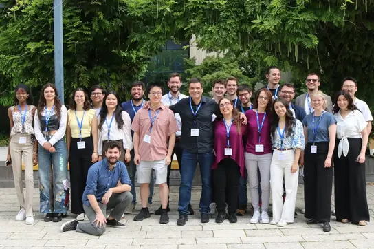
Biomedical Physics in Radiation Oncology - Group Photo
FLASH 2024 Workshop Group Photo
FLASH Workshop 2025
Team
Short description text to introduce the team members.
- Show profile
Prof. Dr. Joao Seco
Head of Division of Biomedical Physics in Radiation Oncology
- Show profile
Lisa Alborghetti
PhD Student
- Show profile

Mark Arndt
PhD Student
- Show profile

Mariana Bras
PhD student
-
Viktoriia Bulanova
- Show profile

Sandra Casula
Administrative Assistant for Divisions E041, E270 and E280
- Show profile
Helena Dilling
Bachelor Student
- Show profile

Ruirui Dong
PhD Student
- Show profile
Diogo Engrácia
PhD Student
- Show profile

Rafael Espinhosa Cepeda Lopes
Master Student
- Show profile

Daniel Garcia
Postdoctoral Fellow
- Show profile
Cátia Filipa Gouveia Rosa
PhD Student
- Show profile
Emil Hain
Bachelor Student
- Show profile
Sarah Hasan
Master Student
- Show profile

Niklas Jung
Bachelor Student
-
Dr. Clarence King
- Show profile
Konstantinos Koritsidis
Master Student
- Show profile
Simon Kubillus
Bachelor Student
- Show profile
Patrick Kudis
Master Student
-
Pinelopi Violeta Ladopoulou
- Show profile
Joana Leitao
PhD Student
- Show profile
Art Lindner
Bachelor Student
- Show profile

Dr. Styliani Logotheti
Postdoctoral Fellow
- Show profile
Piet Meiners
Bachelor Student
- Show profile

Hugo Filipe Meles Freitas
Postdoctoral Fellow
-
Alba Meneses Felipe
-
Miguel Molina-Hernandez
-
Esmaeil Nobakht
- Show profile
Ömer Nuhoglu
Master Student
- Show profile

Joao Pedro Oliveira Lourenco
PhD Student
- Show profile

Dr. Francesca Pagliari
Deputy Head of Division of Biomedical Physics in Radiation Oncology
- Show profile

Dr. Alexander Pryanichnikov
Postdoctoral Fellow
-
Sandra Raquel Quitério Carreira
- Show profile
Kurt Schnepp
Bachelor Student
- Show profile

Mats Stauske
Master Student
- Show profile

Chiara Tagliavini
Master Student
- Show profile
Elpida Theodoridou
PhD Student
Projects
BONEOSCOPY: Novel In-vivo Spectroscopy Analysis Technology for Particle Therapy
A novel technology for in vivo spectroscopic analysis of tissue during particle beam therapy is being developed as part of the European Innovation Council's (EIC) Pathfinder Open funding program. Metastatic bone cancer is an incurable disease and one of the most complex cancers to treat. Due to the high dose, tumor imaging is currently performed at the beginning and end of standard particle radiation therapy (PRT), making personalized treatment difficult. The primary goal of the Pathfinder Open: BoneOscopy project is to develop a radically new technology to enable informed medical decisions by monitoring bone cancer on a daily basis during PRT. (https://accelopment.com/projects/boneoscopy/)
IFIGENIA: New Linac for Nuclear Medicine and Molecular Imaging
Nuclear medicine and molecular imaging are widely used to diagnose and treat a wide range of diseases, including cancer, cardiovascular disease and brain disorders such as Alzheimer's and Parkinson's disease. However, the number of nuclear medicine procedures in Europe is significantly lower because most European countries lack the specialized equipment needed to produce the radioisotopes. Within the framework of the Horizon European Research Executive Agency (REA), funding has been received to develop a center of excellence (Excellence Hub) in South Eastern Europe (Greece, Slovenia, Bosnia and Herzegovina and Cyprus) to develop a novel accelerator dedicated to securing a production platform for a wide range of radioisotopes. A LINear ACcelerator (LINAC) provides a compact, cost-effective and environmentally friendly option that can be located in close proximity to hospitals. The tunability of LINACs allows energy levels, currents and targets to be adjusted, enabling the production of a wide range of radioisotopes. In particular, a similar facility, called NUSANO[5], is being built in West Valley City, Utah, USA, and will be operational in 2025. The novel center of excellence is called "IFIGENIA".
FLASH Zebra: Investigation of Oxygen Depletion Hypothesis
The project focuses on achieving a scientific breakthrough in determining the role of oxygen at the FLASH effect. It is well known that irradiation with high dose rates (>40 Gy/s) causes a protective effect of healthy tissue while maintaining tumor control (FLASH effect). However, the mechanisms contributing to this effect are yet to be understood. Starting from the oxygen partial pressure (pO2), a critical parameter for the prevalence of the FLASH effect, this project aims to design in-vivo oxygen measurements during conventional and FLASH radiation treatment and thus to correlate oxygen distribution and depletion with biological endpoints in zebrafish embryos (ZFE).
PROMPT FLASH: Developing real-time dosimetry for in-vivo assessment during FLASH
FLASH radiotherapy demonstrates a striking biological sparing effect of normal tissues while keeping similar anti-tumor efficacy, termed the “FLASH effect”. The measurement of dose at ultra-high-dose-rates or FLASH rates is complex as dosimeters may be susceptible to saturation at ultra-high dose rates. An in-vivo method of verifying the delivered FLASH dose is currently not available. It is vital to guarantee the accuracy of the delivered dose during FLASH. The current proposal focuses on a new method for verifying delivered FLASH dose in-vivo using prompt gamma x-ray spectroscopic (PGXS) analysis of irradiated tissue, which uses Gadolinium-Based Contrast Agents (GBCA) to amplify gamma/X-ray emission. A graphite calorimetry methodology will be used as absolute dose reference.
gROS MC-FLASH: Developing novel Monte Carlo tool for FLASH
The application of ultra-high dose rates (>40 Gy/s) or FLASH radiotherapy (FLASH-RT) has gained significant interest during the last years, when compared to conv-RT (given at 2 Gy/min) due to more than 50% reduction in toxicity in healthy lung tissue of mice. Despite extensive development of the Monte Carlo (MC) codes to
FLASH-RT, the codes struggle to model accurately the chemical impact of the different pulse structures. The Monte Carlo codes have not been capable of explaining why the H2O2 yield is lower in FLASH beams relative to conv-RT beams in pure water. The goal of the project is to develop and customize gROS, a new MC tool capable of simulating the production and diffusion of ROS for various dose-rate and pulse structure deliveries of radiation dose. The gROS MC tool
will be validated via measurements done in water (H2O) of ROS, H2O2 and O2 consumption, for the various pulse structures and dose rates. The gROS Monte Carlo will be tested at the Marburg Ion-Beam Therapy Center (MIT) facility for various cell types (cancer and healthy) and for particles: electrons, protons and carbon ions. The key objectives are (1) To develop a gROS Monte Carlo tool capable of simulating production and diffusion of ROS and H2O2 for various low
(conv-RT) and ultra-high dose rate (FLASHRT) deliveries, for different pulse structures and at different oxygen concentrations within the medium; (2) To validate the gROS Monte Carlo tool based on measurements in H2O of O2 consumption and H2O2 (ROS) production for various pulse structures and at different oxygen concentrations within the medium; (3) To assess gROS estimate of ROS (H2O2) and O2 consumption by the radiation in an in-vitro 2D model of cancer and healthy cells irradiated with electrons, protons and carbon ions.
PrecisionXray (PXI) FLASH Shutter: Development of a table-top FLASH shutter system
A novel shutter is being developed jointly with PrecisionXray Inc, for allowing table-top FLASH conditions for Xray beams.
HELIOS: Development of Novel Particle Imaging Device for mixed He-C beam Imaging
Development of a novel range guided radiotherapy (RGRT) prototype to reduce toxicity in carbon ion therapy of non-small cell lung carcinoma (NSCLC), by invivo monitoring of the delivered dose to a moving tumor. The RGRT approach is based on a carbon beam with mixed-in helium ions, resulting in helium ions exiting the patient due to their longer range at same energy per nucleon. The exiting helium ions will allow for in-vivo and real-time monitoring of the carbon Bragg peak within the patient with 2mm accuracy, reducing the amount of healthy tissue that is irradiated. This will reduce patient toxicity and allow for improved tumor control.
Selected Publications
Jansen J, Knoll J, Beyreuther E, Pawelke J, Skuza R, Hanley R, Brons S, Pagliari F, Seco J.
Zhang T, Stengl C, Derksen L, Palskis K, Koritsidis K, Zink K, Adeberg S, Major G, Weishaar D, Theiß U, Jin J, Spadea MF, Theodoridou E, Hesser J, Baumann KS, Seco J.
Dal Bello R, Becher T, Fuss MC, Krämer M, Seco J
Zhang T, García-Calderón D, Molina-Hernández M, Leitão J, Hesser J, Seco J.
Heidelberg Institute for Radiation Oncology (HIRO)
Our division is member of the Heidelberg Institute for Radiation Oncology (HIRO).
Get in touch with us


