Abdomen Phantom
Stefan Schaller
The aim of this phantom is to study the properties of cone-beam image reconstruction algorithms based on simulations. A typical diagnostic problem in the abdominal region is the detection of liver lesions. Demands on high contrast resolution are moderate, but artifacts occasionally bury anatomical low-contrast structures.
Phantom Description
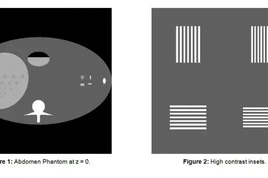
Our phantom consists of a main body, modeled by an elliptical cylinder 40cm wide and 24cm thick. An ellipsoid of dimensions 14cm x 16cm x 20cm models the liver. We have put low-contrast lesions into the liver. The contrast is 10 HU. The lesions are represented by spheres with diameters 2mm, 3mm, 4mm, 5mm, 6mm, 8mm, 10mm and 12mm respectively. Lesions are placed both in the plane at z=0 and in the plane at z=3cm. There are 5 vertebrae at z=-10cm, -5cm, 0cm, 5cm and 10cm. Particular problems arise at long straight contrast edges, such as liquid/air surfaces. To be able to study that particular problem, we have put into the phantom a hollow object, which is partly filled by a liquid. Another major cause of artifacts in spiral CT scanning are high contrast structures tilted with respect to the axis of rotation. Our phantom therefore contains two ribs on the left and right, tilted by 60 degrees with respect to the longitudinal axis. The phantom also contains high contrast insets that allow us to verify that a desired high contrast resolution has been achieved. Resolutions are 10 lp/cm and 12 lp/cm both in azimuthal and radial direction respectively. (Note: The vertebrae are defined in a separate include file.)
Files
- Phantom Definition
- Zip
- Syntax Explanation
