Translational Molecular Imaging in Oncologic Therapy Monitoring
- Imaging and Radiooncology
- DKFZ Hector Cancer Institute
- Clinical Cooperation Unit
- Junior Research Group

Priv. Doz. Dr. Freba Grawe
Group leader
Management of cancer patients can be challenging, especially when it comes to selecting the most suitable treatment that promises the best success with minimal side effects. To monitor treatment success, regular follow-up checks are conducted.

Our Research
Molecular imaging plays a crucial role in this process. Radioactive substances are administered to the patient to visualize biological processes non-invasively using Positron Emission Tomography (PET). The combination of PET with Computed Tomography (PET/CT) or Magnetic Resonance Imaging (PET/MRI) allows us to capture functional and morphological information simultaneously.
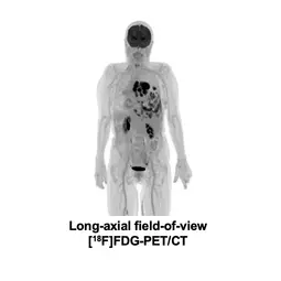
Our goal is to harness the capabilities of molecular imaging, especially PET, to translate research results into clinical practice. In our clinical cooperation unit, experts from various disciplines, including nuclear medicine physicians, radiologists, radiopharmacists, and medical physicists work together on the development and establishment of innovative PET tracers using cutting-edge technology such as the long-axial field-of-view PET/CT, PET/MRI, and photon-counting CT to advance personalized oncology. We aim to improve the early detection of cancer and precise prediction of therapy response for our patients. Our work represents a significant step toward precision medicine for oncology patients.
Projects

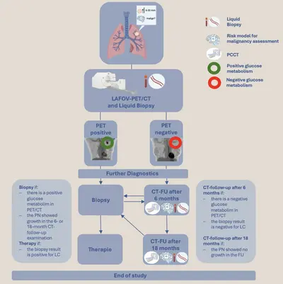
The aim of the PRINCIPLE study is to improve the early and precise characterization of pulmonary nodules through innovative, multimodal diagnostics.
State-of-the-art imaging technologies, such as the "long-axial field-of-view Positron Emission Tomography/Computed Tomography" (LAFOV-PET/CT) and Photon-Counting Computed Tomography (PCCT), are combined with innovative molecular genetic analyses from both liquid and tissue biopsies. Artificial intelligence supports the detection of pulmonary nodules, image evaluation, and integration of multimodal examinations to precisely predict the malignancy of pulmonary nodules. The study overview is presented in Figure 1.
Patients with a pulmonary nodule between 6 and 30 mm without known cancer diagnosis in the past five years are eligible to participate in the study after consultation with a pulmonologist.

Immunotherapy has revolutionized the treatment landscape for lung cancer. However, precise and early assessment of treatment response and survival is inadequately possible with established methods such as computed tomography.
The SPARKLE study investigates the value of innovative hybrid imaging using LAFOV-PET/CT to visualize activated fibroblasts and tumor metabolism for therapy monitoring in patients with lung cancer. Complementary to this, molecular analyses from both liquid and tissue biopsies provide dynamic insights into tumor evolution and treatment response.
This study combines state-of-the-art imaging technologies with advanced molecular analysis methods to better understand and predict the response to immunotherapy in lung cancer.
Treatment with antibodies and antibody-drug conjugates (ADCs) has shown significant efficacy and improved survival in patients with various tumors. However, drug resistance, tumor heterogeneity, and changes in target molecule expression over time remain major challenges.
The EPIC and THE-LUNG studies investigate the role of innovative radiolabeled antibodies and antibody-drug conjugates (ADCs) for non-invasive assessment of the entire tumor compared to current biopsy-based methods and precise therapy monitoring. These studies utilize advanced imaging technologies to provide comprehensive insights into tumor characteristics, potentially helping to select patients most likely to respond to treatment.
Fundings and Research Cooperations
The PRINCIPLE study is funded by the Hector Seed Funding 2024.
The PRINCIPLE study is supported by a research cooperation with Optellum.
The EPIC and THE-LUNG studies are funded by the DKFZ Management Board.

Team
-

Priv. Doz. Dr. Freba Grawe
Group leader
-

Dr. Anna Hinterberger
Deputy group leader
-

Letizia Vella
MTRA Manager
-

Sergej Lossew
MTRA
-
Marie Feline Prochiner
Scientific Associate
Selected Publications
Grawe F et al.
Grawe F et al.
Grawe F et al.
Fabritius MP et al.
Get in touch with us

