Core Facility Small Animal Imaging

Dr. Manfred Jugold
Head of the Small Animal Imaging Core Facility
The Core Facility Small Animal Imaging (SAI), led by Dr. Manfred Jugold, provides comprehensive services for the non-invasive characterization of small animal pathologies and therapeutic effects for internal groups at DKFZ. We assist in documenting pathologies, therapy monitoring, and measuring quantitative and functional tissue parameters. Meaningful images obtained from various modalities can significantly enhance presentations and publications.

Services
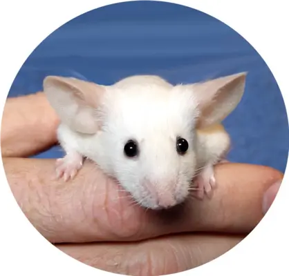
The Small Animal Imaging Unit offers a wide range of services in the field of modern non-invasive in vivo imaging techniques for all internal groups and their collaborators on campus. Our services include project planning, assistance in the formulation of animal research applications, execution of all imaging experiments, storage and analysis of raw data, evaluation of results, and support in the preparation of publications. In close collaboration, we work with you to develop an optimal experimental plan, ensuring that highly focused approaches yield the maximum amount of significant information. Your requirements will guide the selection of modalities and methods. We ensure meticulous execution and professional analysis to meet your expectations.
Please do not hesitate to contact us for a preliminary consultation.
Instrumentation
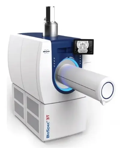
This high-field magnetic resonance imaging (MRI) technique provides detailed images of soft tissues, making it invaluable in tracking tumor development and assessing treatment response in preclinical cancer models. The enhanced resolution allows researchers to visualize anatomical structures and identify changes associated with tumor growth.
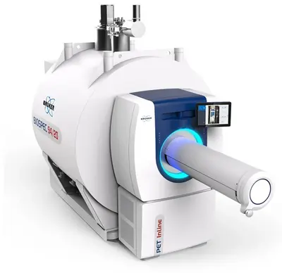
As a higher-resolution variant than the 3 Tesla MRI, the 9.4 Tesla MRI offers even greater detail, particularly useful for studying small animal models of cancer. Its superior sensitivity enables the detection of subtle morphological changes and early-stage tumors that might be missed by lower field strengths.
Micro-computed tomography (µCT) is significant in applied cancer research with laboratory animals as it provides high-resolution, three-dimensional imaging of tumor morphology and vascularization. This non-invasive technique allows for longitudinal studies, enabling researchers to monitor tumor growth and response to therapies over time. Additionally, µCT facilitates the assessment of bone involvement and other structural changes associated with cancer progression.
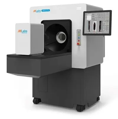
Positron Emission Tomography (PET) is a functional imaging technique that assesses metabolic activity within tissues by detecting gamma rays emitted from radiotracers, often used to evaluate tumor metabolism and proliferation in vivo. This method is critical for monitoring response to therapies and understanding disease progression.

Single Photon Emission Computed Tomography (SPECT): Similar to PET, SPECT provides functional information about blood flow and metabolic activity but uses different radiopharmaceuticals with longer half life times to produce three-dimensional images. It plays an essential role in evaluating treatment effects on tumor viability and studying physiological processes related to cancer.
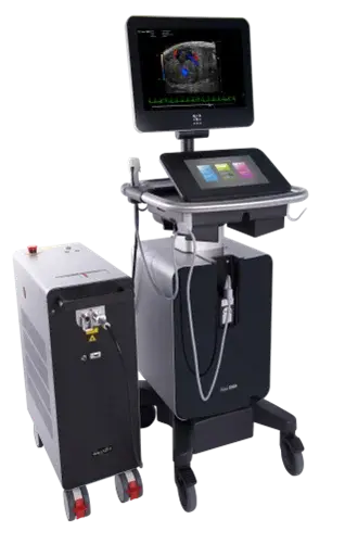
This non-invasive imaging modality uses high-frequency sound waves to create real-time images of internal body structures, including tumors in live animals. Ultrasound is particularly beneficial for guiding biopsies or interventions due to its safety profile and ability for immediate imaging feedback. Ultrasound, allows for the assessment of blood flow by measuring frequency shifts of sound waves reflecting off moving blood cells. This enables quantitative analysis of parameters like blood velocity and perfusion in tumors. Such measurements are crucial for studying tumor angiogenesis and evaluating treatment responses.
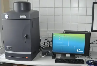
Optical imaging techniques, such as bioluminescence and fluorescence imaging, allow researchers to visualize tumors and biological processes at the cellular level using light-emitting reporter genes or fluorescent markers. This method is advantageous for tracking tumor growth, metastasis, and the effectiveness of treatments over time in live animal models.
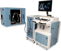
Combining optical and ultrasoundimaging, photoacoustic imaging provides high-resolution images by detecting ultrasonic waves generated from absorbed light in tissues. This technique is especially useful in cancer research for visualizing tumor vasculature and assessing oxygen saturation, which can help in evaluating tumor microenvironments and their responses to therapies.
Each modality offers a variety of application possibilities. Please feel free to reach out for a detailed consultation.
Team

Publications
Authorships
under construction
Acknowledgements
under construction
Get in touch with us

