Bone marrow working group
Imaging in monoclonal plasma cell diseases

[autotranslated from German content]
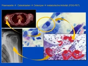
The haemato-oncological imaging working group deals with the possibilities of non-invasive imaging of haematopoietic tissues and the lymphatic system. In particular, patients with hematologic diseases are examined using conventional and functional cross-sectional imaging techniques. In particular, magnetic resonance imaging and computed tomography techniques are used. The data obtained are then correlated with serological, genetic and clinical data. In particular, we are investigating whether dynamic contrast-enhanced MRI (DCE-MRI) and diffusion-weighted imaging (DWI) can help to predict the course of the disease in patients with newly diagnosed multiple myeloma and monoclonal gammopathy of undetermined significance (MGUS). In addition, analyses of large patient collectives that have been examined using standard imaging procedures such as whole-body MRI and whole-body CT as part of routine clinical practice are being carried out. Affected bone marrow areas are identified and monitored over the course of the disease. In particular, the transition from early stages of the disease that do not require treatment to advanced stages will be investigated. In addition, residual findings of the disease after therapy are investigated for their prognostic significance.
Bone marrow and chemotherapy
In contrast to a bone marrow biopsy, MR imaging of the bone marrow, especially with the whole-body technique, provides a sensitive and non-invasive method to visualize the marrow as a whole organ, whereas histology reveals more information about the cell composition at the local level, but its representativeness is highly dependent on the sampling site. Toxic effects on the bone marrow, which can be caused by non-cell-specific chemotherapies or other drugs, significantly influence co-morbidity and survival.
Bone marrow edema, hypo- or aplasia, reconversion and necrosis are the most common changes associated with a wide range of chemotherapeutic agents and their additives.
Knowledge of short- and long-term effects of chemotherapy on the normal bone marrow is essential for the correct interpretation of bone marrow signaling changes in cancer patients. In addition to conventional spin-echo sequences, we are investigating newer MR techniques (e.g. chemical shift imaging, diffusion-weighted imaging, dynamic contrast-enhanced MRI and spectroscopy), all of which support the interpretation of benign and malignant bone marrow changes.
Cooperation partner

Medical Clinic V, Hematology, Oncology and Rheumatology, Heidelberg University Prof. Dr. H. Goldschmidt, Dr. Dirk Hose, Prof. Dr. Jens Hillengass, Dr. Marc Raab
Institute of Pathology, Heidelberg University
Dr. M Andrulis
National Cancer Institute, Bethesda, MA, USA
Prof. Ola Landgren MD, PhD
Division of Medical Physics in Radiation Therapy
Prof. Dr. R. Bendl, Ms. Andrea Fränzle
Selected publications
Infiltration patterns in monoclonal plasma cell disorders: correlation of magnetic resonance imaging with matched bone marrow histology.
Andrulis M, Bäuerle T, Goldschmidt H, Delorme S, Landgren O, Schirmacher P, Hillengass J.Eur J Radiol. 2014 Jun;83(6):970-4. doi: 10.1016/j.ejrad.2014.03.005. Epub 2014 Mar 22.
Imaging of Complications and Toxicity following Tumor Therapy: Bone Marrow and Chemotherapy; Medical Radiology - Diagnostic Imaging, Jobke B., Springer Verlag, 2015 (online first, in print)
Hillengass J, Ayyaz S, Kilk K, Weber MA, Hielscher T, Shah R, Hose D, Delorme S, Goldschmidt H, Neben K. Changes in magnetic resonance imaging before and after autologous stem cell transplantation correlate with response and survival in multiple myeloma. Haematologica. 2012 Jun 11.
Hillengass J, Bäuerle T, Bartl R, Andrulis M, McClanahan F, Laun FB, Zechmann CM, Shah R, Wagner-Gund B, Simon D, Heiss C, Neben K, Ho AD, Schlemmer HP, Goldschmidt H, Delorme S, Stieltjes B. Diffusion-weighted imaging for non-invasive and quantitative monitoring of bone marrow infiltration in patients with monoclonal plasma cell disease: a comparative study with histology. Br J Haematol. 2011 Jun;153(6):721-8
Hillengass J, Fechtner K, Weber MA, Bäuerle T, Ayyaz S, Heiss C, Hielscher T, Moehler TM, Egerer G, Neben K, Ho AD, Kauczor HU, Delorme S, Goldschmidt H. Prognostic significance of focal lesions in whole-body magnetic resonance imaging in patients with asymptomatic multiple myeloma. Journal of Clinical Oncology 2010 Mar 20;28(9):1606-10