Ultrasound (US)
Ultrasound (syn. sonography; abbreviation US) - a gentle examination using sound waves

An ultrasound examination is a non-invasive and informative examination using ultrasound waves. Like computer tomography, it is a cross-sectional imaging procedure. This method can produce moving images in real time, particularly of various abdominal organs such as the spleen, liver, pancreas and kidneys. However, other organs such as the thyroid gland, parathyroid glands, the soft tissues of the neck or the female breast can also be examined very easily and with little stress for the patient. As ultrasound examinations do not involve X-rays, they can also be used in the event of pregnancy. If pregnancy cannot be ruled out, this is not an obstacle to the examination.
Ultrasound examination at the DKFZ - information on the procedure and valuable tips for the patient
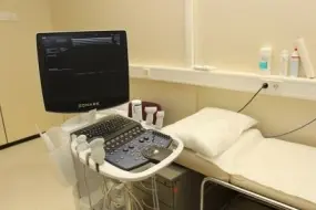
Many different ultrasound examinations are carried out at the DKFZ.
Air-filled intestinal loops can make it difficult to assess certain abdominal organs (e.g. pancreas). It is therefore important that no flatulent meals are consumed before the examination. It is important to come to certain examinations of the abdominal organs on an empty stomach. Please consult your referring doctor or our registration department.
Depending on the question, the area of the body to be examined (neck, abdomen) must be completely undressed. In order to create good sound conditions for the examining doctor, you will lie in a darkened room; you will usually lie on your back; it may be necessary to reposition the patient during the examination. For certain organs, you may have to turn to the side or stretch your head. To create good sound conditions for the examiner, ultrasound gel is used to prevent the reflection of sound waves in the air. The ultrasound probe must occasionally be pressed harder. This can sometimes be uncomfortable - please inform the examining doctor immediately.
The examination itself takes approx. 15-20 minutes. After the examination, you can get dressed again at your leisure and will usually be asked to take another short seat in the waiting room. If the ultrasound examination with contrast medium was performed, the venous access in your arm will now be removed again. After consultation with the responsible senior physician, the ultrasound images will be discussed and then evaluated by two examiners; the findings will then be sent to your referring physician.
Are contrast agents also used in ultrasound examinations?
Yes, for certain questions it may be necessary to inject a contrast agent into the bloodstream during ultrasound, although this is rarely the case. For this purpose, a so-called brown needle (small plastic tube) will be inserted into a vein before the examination begins. During the examination, this contrast medium is injected by hand. This usually serves to improve the contrasting of vessels and organs. It enables a precise distinction to be made between areas with good and poor blood flow. These are substances that are tolerated by the body without any problems. Therefore, no side effects - apart from occasional pain at the injection site - are to be expected. However, the contrast medium should not be used in the case of certain metabolic diseases (galactosemia), severe heart failure, recent heart attacks and severe (obstructive) lung diseases.