Cookie Settings

We use cookies to optimize our website. These include cookies that are necessary for the operation of the site, as well as those that are only used for anonymous statistic. You can decide for yourself which categories you want to allow. Further information can be found in our data privacy protection .

Essential

These cookies are necessary to run the core functionalities of this website and cannot be disabled.

Name Webedition CMS
Purpose This cookie is required by the CMS (Content Management System) Webedition for the system to function correctly. Typically, this cookie is deleted when the browser is closed.
External media

Content from external media platforms is blocked by default. If cookies from external media are accepted, access to this content no longer requires manual consent.

Name YouTube
Purpose Show YouTube content
Name Twitter
Purpose activate Twitter Feeds

Double imaging of prostate cancer

No. 35c | 26/06/2017 | by Koh

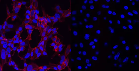
Scientists from the German Cancer Research Center (DKFZ) are developing a radiopharmaceutical that visualizes prostate cancer in two different ways: Radioactive labeling facilitates detection of a tumor or its metastases in the body. At the same time, a coupled fluorescent dye helps surgeons identify and safely remove cancer tissue during surgery.

© Baranski/DKFZ

The so-called bimodal radiopharmaceutical binds to prostate-specific membrane antigen (PSMA). PSMA is a surface protein that is normally present on healthy prostate cells, but is found at much higher levels on prostate cancer cells. It is barely found in the rest of the body. Therefore, PSMA is an ideal target for diagnostic purposes as well as targeted therapies against prostate cancer.

Radioactive labeling of the bimodal pharmaceutical as a PET tracer makes it possible to locate the tumor and its metastases using a combination of positron emission tomography (PET) and computerized tomography (CT) or magnetic resonance imaging (MRI), respectively. This non-invasive imaging can also be used for surgical planning.

During surgery, the fluorescent dye that is coupled to the pharmaceutical helps physicians differentiate between malignant and healthy tissue, thus facilitating very precise removal of tumor tissue. This combined approach of imaging and therapy will substantially increase the effectiveness of surgical interventions.

© Baranski/DKFZ

Ann-Christin Baranski from the German Cancer Research Center (Deutsches Krebsforschungszentrum, DKFZ) was honored with the 2017 Image of the Year Award of the Society of Nuclear Medicine and Molecular Imaging (SNMMI) at its Annual Meeting.SNMMI awards the prestigious distinction for the most promising advances in the field of nuclear medicine and nuclear imaging.

The project led by Matthias Eder, German Cancer Consortium (DKTK) Freiburg, and Klaus Kopka, DKFZ, has been supported since 2016 by the Federal Ministry of Education and Research (BMBF) as part of its VIP+ (Validation of the technological and social innovation potential of scientific research) funding program. This initiative helps scientists examine and prove the innovation potential and possible applications of their research results.

The researchers' goal is a first clinical application of the bimodal radiopharmaceutical that can be used in a combined approach for nuclear-medical imaging (PET/MRT or PET/CT) and for subsequent intra-operative fluorescence-guided navigation during robot-assisted removal of prostate cancer and its metastases.

Please find more information at:

http://www.snmmi.org/NewsPublications/NewsDetail.aspx?ItemNumber=24411

http://www.auntminnie.com/index.aspx?sec=sup&sub=mol&pag=dis&ItemID=117606
With more than 3,000 employees, the German Cancer Research Center (Deutsches Krebsforschungszentrum, DKFZ) is Germany’s largest biomedical research institute. DKFZ scientists identify cancer risk factors, investigate how cancer progresses and develop new cancer prevention strategies. They are also developing new methods to diagnose tumors more precisely and treat cancer patients more successfully. The DKFZ's Cancer Information Service (KID) provides patients, interested citizens and experts with individual answers to questions relating to cancer.

To transfer promising approaches from cancer research to the clinic and thus improve the prognosis of cancer patients, the DKFZ cooperates with excellent research institutions and university hospitals throughout Germany:

  • National Center for Tumor Diseases (NCT, 6 sites)
  • German Cancer Consortium (DKTK, 8 sites)
  • Hopp Children's Cancer Center (KiTZ) Heidelberg
  • Helmholtz Institute for Translational Oncology (HI-TRON Mainz) - A Helmholtz Institute of the DKFZ
  • DKFZ-Hector Cancer Institute at the University Medical Center Mannheim
  • National Cancer Prevention Center (jointly with German Cancer Aid)
The DKFZ is 90 percent financed by the Federal Ministry of Education and Research and 10 percent by the state of Baden-Württemberg. The DKFZ is a member of the Helmholtz Association of German Research Centers.

RSS-Feed

Subscribe to our RSS-Feed.

to top
powered by webEdition CMS