A couple of years ago, London taxi drivers produced very impressive proof of the key role which the hippocampus plays for spatial orientation. Studying the brains of taxi drivers, British neurologists were able to detect characteristic volume changes in this part of the brain, which were found to correlate with the amount of time spent as a taxi driver.
Researchers have studied how the hippocampus works and what functions it has not only in taxi drivers but also in mice. From these studies, neuroscientists know that most signals that are to reach the hippocampus have to pass a special structure of the cortex called entorhinal cortex, which is a kind of bottleneck on the way to the hippocampus. These two brain regions communicate closely with each other and are directly interconnected via a multitude of long neuronal projections. “However, only the presence of excitatory nerve fibres between these two areas of the brain has been known so far," said Professor Hannah Monyer, commenting on her recent findings. The neuroscientist leads a collaborative department of the German Cancer Research Center (DKFZ), Heidelberg University and Heidelberg University Hospital. “Now we have been able to show that there are also inhibitory neurons, which release the GABA neurotransmitter, forming direct connections between the two structures and, thus, contributing to the interactions of the two brain areas."
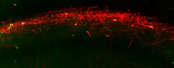
Using a new detection method, the researchers were able to image the individual neuronal connections in the brain and study their functions in detail at the same time: In brain tissue of mice they specifically introduced a light-sensitive fluorescent protein into the inhibitory GABA neurons. With the aid of the fluorescent marker it was possible to visualize the exact course of the long projections of the neurons between the two brain areas. This method also made it possible to identify the target cells of the new direct connections; for the most part, these are what are called inhibitory interneurons. This type of neurons forms local connections between hundreds of neighboring neurons, thus setting the pace in whole areas of the brain.
In the next step, the group studied what happens in the downstream interneurons when a signal is transmitted via the long-range connection: The investigators activated the introduced light-sensitive protein by laser pulses, thus specifically inducing electrical discharges in individual long-range neurons. The researchers were able to measure that the activity of target cells is being inhibited at this moment.
Even though the long-range inhibitory neurons are just a minority among the total population of neurons, the effect of their activation is immense. This is no surprise considering that their target cells, interneurons, act like conductors synchronizing whole orchestras of nerve cells. Figuratively speaking, the newly discovered long-range inhibitory neurons are supersynchronizers of conductor cells which lead their own subordinated orchestras.
The researchers in Monyer‘s team have already started verifying their findings obtained in tissue sections in living mice. „With every new neuronal circuit that we discover and understand we gain a better overall picture of how different areas of our brain are orchestrated. This coordinated interplay of different structures is the physiological basis of learning and memory,“ said Hannah Monyer, putting her research into a wider context.
Sarah Melzer*, Magdalena Michael*, Antonio Caputi*, Marina Eliava, Elke C. Fuchs, Miles Whittington and Hannah Monyer: Long-Range-Projecting GABAergic Neurons Modulate Inhibition in Hippocampus and Entorhinal Cortex. Science 2012, DOI: 10.1126/science.1217139 (* First authors)


