Light Microscopy

Dr. Felix Bestvater
Head
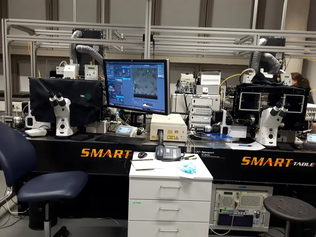
The Light Microscopy Unit provides a wide range of services in the field of modern imaging techniques for all internal groups and their cooperation partners at several locations on campus.

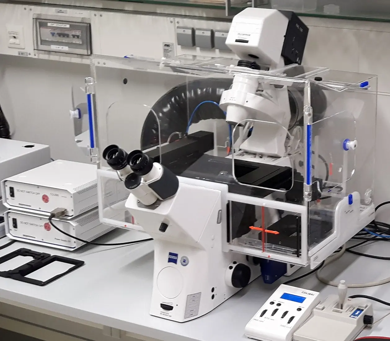
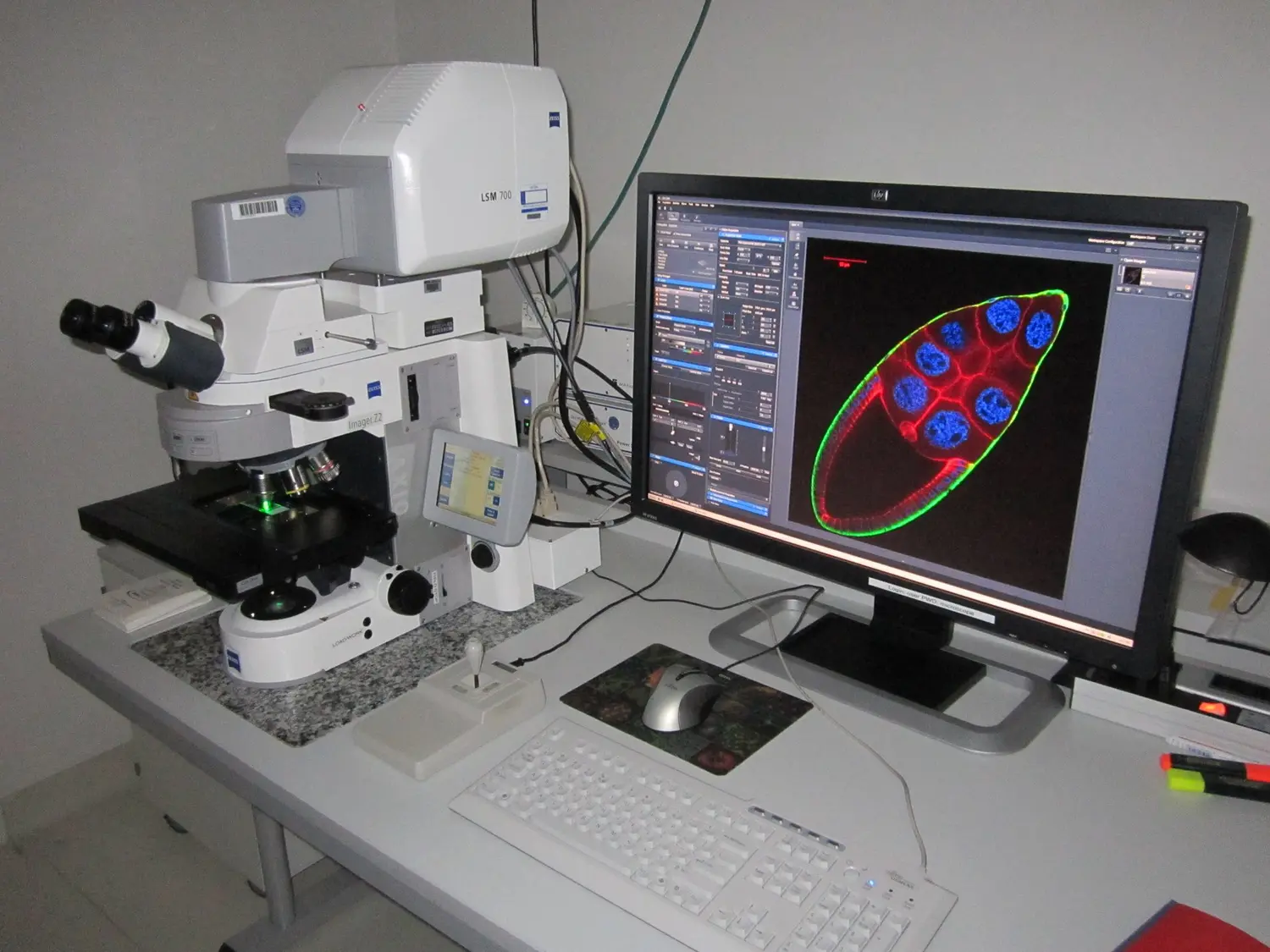
Instrumentation


Technology Development
We provide consultation for special projects and setting up individual protocols for advanced users. We accommodate the increasing demand for automated digital image processing by providing high-end working stations, appropriate software (e.g. ImageJ/Fiji, Arivis, Aivia, Amira, Imaris, ZEN, LAS, Matlab, GraphPad PRISM, SigmaPlot). We provide professional support for image processing algorithm and Fiji/ImageJ macro development to improve reproducibility and efficiency for our users.
Team
-
Dr. Felix Bestvater
Head
-

Manuela Brom
Technician (Microscopy)
-
Dr. Damir Krunic
Scientist
-
Dr. Marko Norman Lampe
Scientist
-
Mahsa Shakouri
Technician (Microscopy)

Publications
Li L, Lopez Perez R, Shehzad K, Jennemann R, Schmidt C, Walle T, Tietz-Dahlfuß A, Grimm E, Kraske JA, Häring P, Barayeu U, Dick TP, Ye L, Braun SA, Hertl M, Worzfeld T, Wiech T, Ji H, Su J, Schneeweiss JM, Liu M, Kommoss K, Heikenwälder M, Zou B, Mücklich S, Steinbrink K, Raker VK, Wu W, Noessner E, Gröne HJ, Nelson PJ, Sandhoff R, Huber PE.
Koch MS, Deo M, Schmidt C, Podolak A, Taranda J, Hoetker MS, Turcan Ş.
Kraske JA, Allers MM, Smirnov A, Lenoir B, Ahmed A, Suarez-Carmona M, Hampel M, Krunic D, Tietz-Dalfuß A, Beikert T, Schneeweiss JM, Brons S, Albrecht D, Trinh T, Liu M, Giese NA, Glowa C, Liermann J, Lopez Perez R, Jäger D, Debus J, Halama N, Huber PE, Walle T.
Siqin S, Rahbari M, Ernst C, Grewe I, Gunst K, Neßling M, Kaden S, Richter K, Häfele L, Feuerbach L, Krunic D, Tessmer C, Hofmann I, Birgin E, Brobeil A, Rahbari N, Heikenwälder M, Bund T.
Imle R, Blösel D, Kommoss FKF, Placke S, Stutheit-Zhao E, Blume C, Lupar D, Schmitt L, Winter C, Wagner L, von Eicke M, Walzer H, Förderer J, Laier S, Hertwig M, Peterziel H, Oehme I, Scheuerman S, Seitz CM, Geyer FH, Cidre-Aranaz F, Grünewald TGP, Vokuhl C, Chudasama P, Scholl C, Schmidt C, Günther P, Sill M, Jones KB, Pfister SM, Autry RJ, Banito A.
Siqin S, Nikitina E, Rahbari M, Ernst C, Krunic D, Birgin E, Tessmer C, Hofmann I, Rahbari N, Bund T.
Jin JX, Fuchslocher F, Carreno-Gonzalez M, Zahnow F, Ceranski AK, Will R, Helm D, Bestvater F, Banito A, Imle R, Ohmura S, Cidre-Aranaz F, Grünewald TGP.
Xu J, Koch J, Schmidt C, Nientiedt M, Neuberger M, Erben P, Michel MS, Rodríguez-Paredes M, Lyko F.
Ultrahigh Dose Rate Helium Ion Beams: Minimizing Brain Tissue Damage while Preserving Tumor Control.
Dokic I, Moustafa M, Tessonnier T, Meister S, Ciamarone F, Akbarpour M, Krunic D, Haberer T, Debus J, Mairani A, Abdollahi A.
Zhou L, Pereiro MT, Li Y, Derigs M, Kuenne C, Hielscher T, Huang W, Kränzlin B, Tian G, Kobayashi K, Lu GN, Roedl K, Schmidt C, Günther S, Looso M, Huber J, Xu Y, Wiech T, Sperhake JP, Wichmann D, Gröne HJ, Worzfeld T.
Berenbrok N, Vargas-Delgado ME, Beitzen-Heineke A, Schmidt C, Gensch V, Loges S, Ben-Batalla I.
Hering M, Madi A, Sandhoff R, Ma S, Wu J, Mieg A, Richter K, Mohr K, Knabe N, Stichling D, Poschet G, Bestvater F, Frank L, Utikal J, Umansky V, Cui G.
Martija AA, Krauß A, Bächle N, Doth L, Christians A, Krunic D, Schneider M, Helm D, Will R, Hartmann C, Herold-Mende C, von Deimling A, Pusch S.
Scherthan H, Geiger B, Ridinger D, Müller J, Riccobono D, Bestvater F, Port M, Hausmann M.
Blanco-Carmona E, Narayanan A, Hernandez I, Nieto JC, Elosua-Bayes M, Sun X, Schmidt C, Pamir N, Özduman K, Herold-Mende C, Pagani F, Cominelli M, Taranda J, Wick W, von Deimling A, Poliani PL, Rehli M, Schlesner M, Heyn H, Turcan Ş.
Nikitina E, Burk-Körner A, Wiesenfarth M, Alwers E, Heide D, Tessmer C, Ernst C, Krunic D, Schrotz-King P, Chang-Claude J, von Winterfeld M, Herpel E, Brobeil A, Brenner H, Heikenwalder M, Hoffmeister M, Kopp-Schneider A, Bund T.
Demirdizen E, Al-Ali R, Narayanan A, Sun X, Varga JP, Steffl B, Brom M, Krunic D, Schmidt C, Schmidt G, Bestvater F, Taranda J, Turcan Ş.
Grimm E, van der Hoeven F, Sardella D, Willig KI, Engel U, Veits N, Engel R, Cavalcanti-Adam EA, Bestvater F, Bordoni L, Jennemann R, Schönig K, Schiessl IM, Sandhoff R.
Schönrock A, Heinzelmann E, Steffl B, Demirdizen E, Narayanan A, Krunic D, Bähr M, Park JW, Schmidt C, Özduman K, Pamir MN, Wick W, Bestvater F, Weichenhan D, Plass C, Taranda J, Mall M, Turcan Ş.
Walle T, Kraske JA, Liao B, Lenoir B, Timke C, von Bohlen Und Halbach E, Tran F, Griebel P, Albrecht D, Ahmed A, Suarez-Carmona M, Jiménez-Sánchez A, Beikert T, Tietz-Dahlfuß A, Menevse AN, Schmidt G, Brom M, Pahl JHW, Antonopoulos W, Miller M, Perez RL, Bestvater F, Giese NA, Beckhove P, Rosenstiel P, Jäger D, Strobel O, Pe'er D, Halama N, Debus J, Cerwenka A, Huber PE.
Dokic I, Meister S, Bojcevski J, Tessonnier T, Walsh D, Knoll M, Mein S, Tang Z, Vogelbacher L, Rittmueller C, Moustafa M, Krunic D, Brons S, Haberer T, Debus J, Mairani A, Abdollahi A.
Gatzweiler C, Ridinger J, Herter S, Gerloff XF, ElHarouni D, Berker Y, Imle R, Schmitt L, Kreth S, Stainczyk S, Ayhan S, Najafi S, Krunic D, Frese K, Meder B, Reuss D, Fiesel P, Schramm K, Blattner-Johnson M, Jones DTW, Banito A, Westermann F, Oppermann S, Milde T, Peterziel H, Witt O, Oehme I.
Cidre-Aranaz F, Li J, Hölting TLB, Orth MF, Imle R, Kutschmann S, Ammirati G, Ceranski K, Carreño-Gonzalez MJ, Kasan M, Marchetto A, Funk CM, Bestvater F, Bersini S, Arrigoni C, Moretti M, Thiel U, Baumhoer D, Sahm F, Pfister SM, Hartmann W, Dirksen U, Romero-Pérez L, Banito A, Ohmura S, Musa J, Kirchner T, Knott MML, Grünewald TGP.
Jennemann R, Volz M, Bestvater F, Schmidt C, Richter K, Kaden S, Müthing J, Gröne HJ, Sandhoff R.
Li J, Ohmura S, Marchetto A, Orth MF, Imle R, Dallmayer M, Musa J, Knott MML, Hölting TLB, Stein S, Funk CM, Sastre A, Alonso J, Bestvater F, Kasan M, Romero-Pérez L, Hartmann W, Ranft A, Banito A, Dirksen U, Kirchner T, Cidre-Aranaz F, Grünewald TGP.
Lan Y, Moustafa M, Knoll M, Xu C, Furkel J, Lazorchak A, Yeung TL, Hasheminasab SM, Jenkins MH, Meister S, Yu H, Schlegel J, Marelli B, Tang Z, Qin G, Klein C, Qi J, Zhou C, Locke G, Krunic D, Derner MG, Schwager C, Fontana RE, Kriegsmann K, Jiang F, Rein K, Kriegsmann M, Debus J, Lo KM, Abdollahi A.
Bartosova M, Ridinger D, Marinovic I, Heigwer J, Zhang C, Levai E, Westhoff JH, Schaefer F, Terjung S, Hildenbrand G, Krunic D, Bestvater F, Hausmann M, Schmitt CP, Zarogiannis SG.
Ohmura S, Marchetto A, Orth MF, Li J, Jabar S, Ranft A, Vinca E, Ceranski K, Carreño-Gonzalez MJ, Romero-Pérez L, Wehweck FS, Musa J, Bestvater F, Knott MML, Hölting TLB, Hartmann W, Dirksen U, Kirchner T, Cidre-Aranaz F, Grünewald TGP.
Popovic ZV, Bestvater F, Krunic D, Krämer BK, Bergner R, Löffler C, Hocher B, Marx A, Porubsky S.
Körholz K, Ridinger J, Krunic D, Najafi S, Gerloff XF, Frese K, Meder B, Peterziel H, Vega-Rubin-de-Celis S, Witt O, Oehme I.
Schwab M, Lohr S, Schneider J, Kaiser M, Krunic D, Helbig D, Géraud C, Angel P.
Wrobel JK, Najafi S, Ayhan S, Gatzweiler C, Krunic D, Ridinger J, Milde T, Westermann F, Peterziel H, Meder B, Distel M, Witt O, Oehme I.
Nowrouzi A, Sertorio MG, Akbarpour M, Knoll M, Krunic D, Kuhar M, Schwager C, Brons S, Debus J, Wells SI, Wells JM, Abdollahi A.
Möhrmann L, Zowada MK, Strakerjahn H, Siegl C, Kopp-Schneider A, Krunic D, Strunk D, Schneider M, Kriegsmann M, Kriegsmann K, Herbst F, Ball CR, Glimm H, Dieter SM.
Hekler A, Utikal JS, Solass W, Schmitt M, Klode J, Schadendorf D, Sondermann W, Franklin C, Bestvater F, Krahl D, von Kalle C, Fröhling S, Brinker TJ.
Hekler A, Utikal JS, Enk AH, Solass W, Schmitt M, Klode J, Schadendorf D, Sondermann W, Franklin C, Bestvater F, Flaig MJ, Krahl D, von Kalle C, Fröhling S, Brinker TJ.
Morace I, Pilz R, Federico G, Jennemann R, Krunic D, Nordström V, von Gerichten J, Marsching C, Schießl IM, Müthing J, Wunder C, Johannes L, Sandhoff R, Gröne HJ.
Meln I, Wolff G, Gajek T, Koddebusch J, Lerch S, Harbrecht L, Hong W, Bayindir-Buchhalter I, Krunic D, Augustin HG, Vegiopoulos A.
Pilarczyk G, Papenfuß F, Bestvater F, Hausmann M.
Even I, Reidenbach S, Schlechter T, Berns N, Herold R, Roth W, Krunic D, Riechmann V, Hofmann I.
Bayindir-Buchhalter I, Wolff G, Lerch S, Sijmonsma T, Schuster M, Gronych J, Billeter AT, Babaei R, Krunic D, Ketscher L, Spielmann N, Hrabe de Angelis M, Ruas JL, Müller-Stich BP, Heikenwalder M, Lichter P, Herzig S, Vegiopoulos A.
Babaei R, Schuster M, Meln I, Lerch S, Ghandour RA, Pisani DF, Bayindir-Buchhalter I, Marx J, Wu S, Schoiswohl G, Billeter AT, Krunic D, Mauer J, Lee YH, Granneman JG, Fischer L, Müller-Stich BP, Amri EZ, Kershaw EE, Heikenwälder M, Herzig S, Vegiopoulos A.
Hildenbrand G, Metzler P, Pilarczyk G, Bobu V, Kriz W, Hosser H, Fleckenstein J, Krufczik M, Bestvater F, Wenz F, Hausmann M.
Popovic ZV, Rabionet M, Jennemann R, Krunic D, Sandhoff R, Gröne HJ, Porubsky S.
Rampoldi F, Brunk F, Bonrouhi M, Federico G, Krunic D, Porubsky S, Gröne HJ, Popovic ZV.
Dokic I, Mairani A, Niklas M, Zimmermann F, Chaudhri N, Krunic D, Tessonnier T, Ferrari A, Parodi K, Jäkel O, Debus J, Haberer T, Abdollahi A.
Dokic I, Niklas M, Zimmermann F, Mairani A, Seidel P, Krunic D, Jäkel O, Debus J, Greilich S, Abdollahi A.
Oleksiuk O, Abba M, Tezcan KC, Schaufler W, Bestvater F, Patil N, Birk U, Hafner M, Altevogt P, Cremer C, Allgayer H.
Osswald M, Jung E, Sahm F, Solecki G, Venkataramani V, Blaes J, Weil S, Horstmann H, Wiestler B, Syed M, Huang L, Ratliff M, Karimian Jazi K, Kurz FT, Schmenger T, Lemke D, Gömmel M, Pauli M, Liao Y, Häring P, Pusch S, Herl V, Steinhäuser C, Krunic D, Jarahian M, Miletic H, Berghoff AS, Griesbeck O, Kalamakis G, Garaschuk O, Preusser M, Weiss S, Liu H, Heiland S, Platten M, Huber PE, Kuner T, von Deimling A, Wick W, Winkler F.
Meister M, Tounsi A, Gaffal E, Bald T, Papatriantafyllou M, Ludwig J, Pougialis G, Bestvater F, Klotz L, Moldenhauer G, Tüting T, Hämmerling GJ, Arnold B, Oelert T.
Get in touch with us