Macromolecular organisation of striated muscle: Sarcomer from murine m. soleus. Insert: cross-section of acto-myosin setup (thick myosin- and thin actin-fibers).
Services
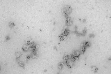
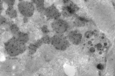
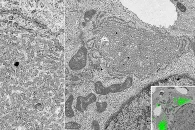
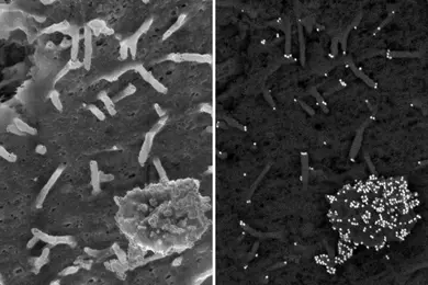
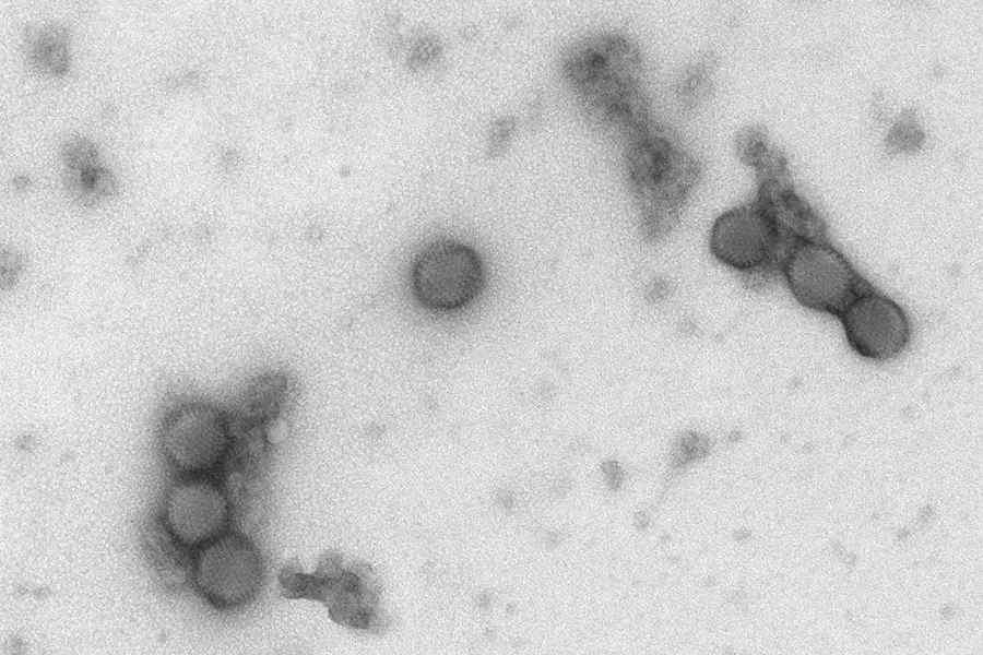
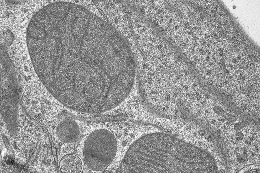
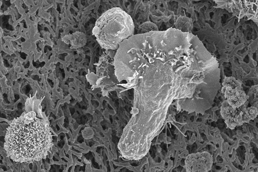
Our service encompasses the design of experimental formats, sample preparation (fixation, embedding, sectioning, contrasting, labelling, carriers, etc.), microscopy, and pre-processing of images. Together with all image data, customers receive the detailed protocol and a report if appropriate. Our portfolio includes negative staining particle-TEM (quality-check of VLP- and EV-preparations), ultrathin sectioning resin-TEM (ultrastructural phenotyping of cells and tissues), as well as SEM (shapes and surface reliefes of cells, organoids and their substrates). Immuno-gold approaches furthermore allow allocation of molecular identity to structures of interest, and CLEM (Correlative Light and Electron Microscopy) to spot sites of interest by fluorescence microscopy. To start a project, contact us to clarify the scientific question and agree on an experimental strategy.
As projects are unique, with respect to sample-formats and structural features of interest, we routinely invest into the development of protocols for sample preparation and imaging.